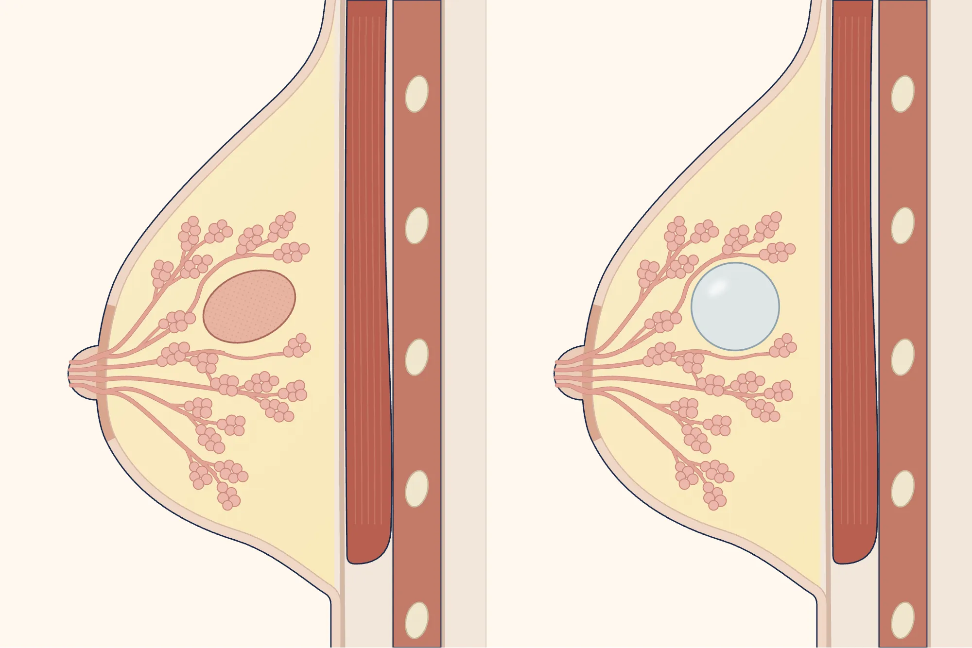
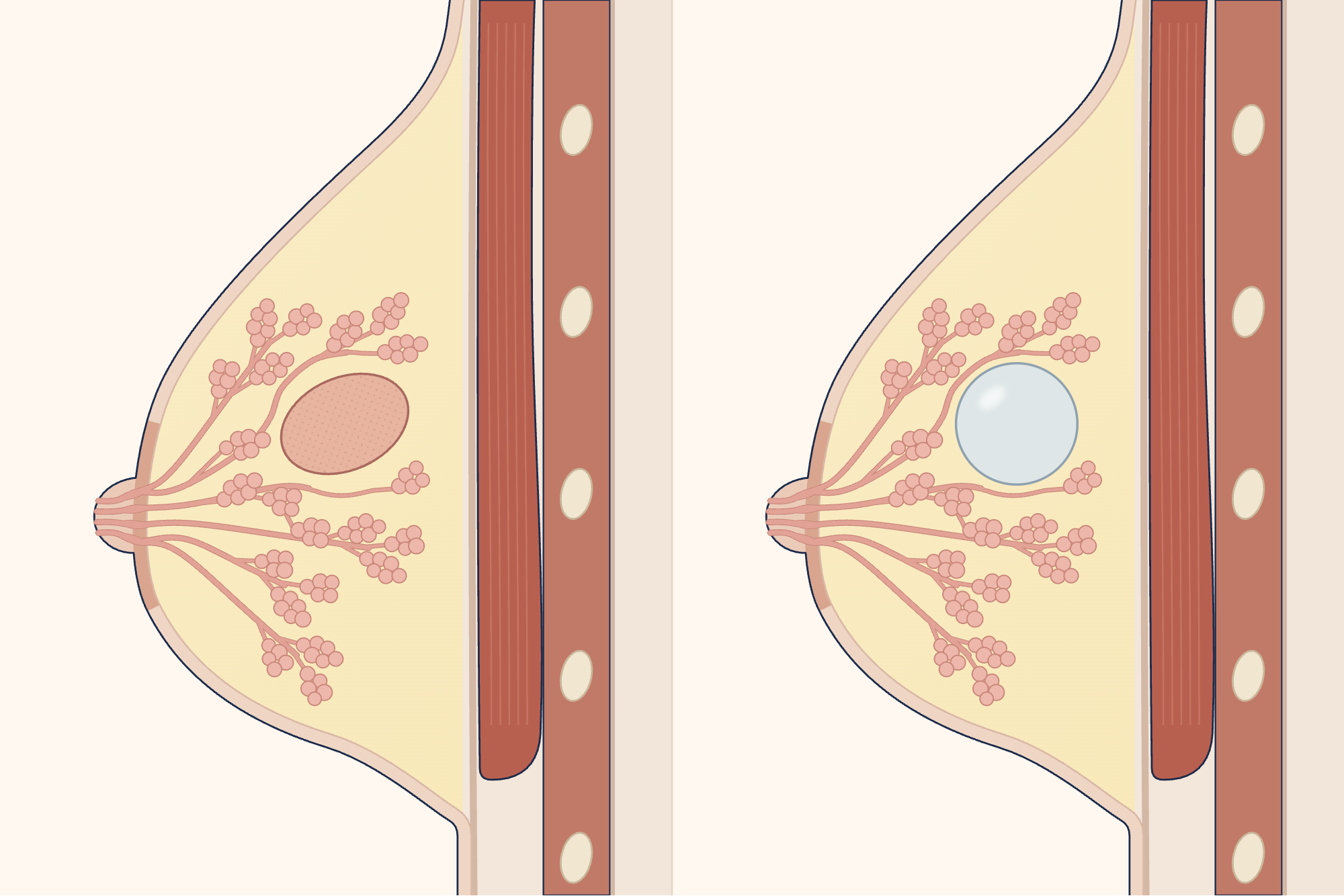

What is a breast lump and what causes it?
Most breast lumps that can be felt are harmless[10]. A lump is often caused by a harmless condition, such as a non-cancerous growth of breast tissue (fibroadenoma) or a collection of fluid (cyst); sometimes, however, a lump is a sign of a serious disease such as breast cancer[10]. Fibroadenomas can occur in around 10 per cent of women and are more common before the menopause[4].
However, a lump that can be felt is the most common symptom of breast cancer[2], so every lump you notice should be examined[10].
What are the symptoms of a breast lump?
A lump may be noticed as a lump or swelling in the breast, chest or armpit[11]. The following changes also require examination, whether they occur with a lump or on their own[11]:
- a change in the size or shape of one or both breasts,
- dimpling of the breast skin (which may look like orange peel),
- a change in the colour of the breast skin: it may look red on lighter skin or darker than usual on darker skin,
- a nipple turning inwards or an eczema-like rash on the nipple,
- discharge from the nipple other than milk leakage during pregnancy and breastfeeding; the discharge may be bloody[11, 12].
These symptoms can also occur without any pain[11]. Nipple discharge may not be anything serious, but sometimes it is a sign of a condition such as a cyst or cancer, so it should be examined[12].
What size in cm is a breast lump dangerous?
Is a painful breast lump dangerous?
A lump should be examined whether it is painful or painless: England’s referral guideline for suspected cancer assesses an unexplained breast lump in the same way whether or not it is painful[3]. If the lump is hard and does not move, see a doctor the same day[13].
Breast pain on its own, without a lump or another change, is not usually a symptom of breast cancer[1, 11]. For pain that spreads over a large part of the breast or comes and goes with the menstrual cycle, imaging beyond the usual screening recommendations is not needed if there are no other suspicious findings[1, 6]. If the pain is in one spot and is unrelated to menstruation, the guidelines differ: the British radiology guideline still does not recommend routine imaging, while the American guideline considers imaging appropriate[1, 6].
Pain that cannot be explained and does not go away should also be assessed by a specialist[5]. See a doctor if you have pain in your breast or armpit and people in your family have had breast or ovarian cancer[11].
How is a breast lump identified?
Lumps are more suspicious if they are hard, do not move because they are attached to the skin or underlying tissue, or cause dimpling of the skin or the nipple to turn inwards; benign lumps, on the other hand, usually move, have well-defined edges and feel soft or rubbery[2]. However, an examination by hand cannot reliably distinguish a benign lump from cancer, or a fluid-filled cyst from a solid lump, even when performed by experienced doctors[2].
Diagnosis is therefore made in three steps: examination, imaging and, when needed, a needle biopsy[1]. These steps are often carried out during the same visit[10].
In women under 40, during pregnancy and while breastfeeding, the first imaging test is an ultrasound; at age 40 and over, a mammogram (breast X-ray) and an ultrasound focused on the lump are performed together[1]. In the American guideline, however, this age threshold is 30 while breastfeeding[8]. If the ultrasound shows a suspicious appearance, a mammogram is also performed[1]. For a lump that can be felt, if a mammogram and ultrasound together show no signs of cancer, studies indicate that the probability of it not being cancer is between 97.4 and 100 per cent; nevertheless, if the examination raises suspicion, a biopsy should be performed even if the imaging is normal[1, 2].
Most solid lumps require a needle biopsy to confirm the diagnosis[1]. Rather than taking cells with a fine needle, taking small pieces of tissue with a thicker needle (core needle biopsy) is recommended because the result is more accurate[1]. The procedure is performed with the area numbed and usually takes 15–20 minutes[5]. The risks are low; very rarely, the membrane surrounding the lung may be punctured[5]. Results are often given on the same day; you may need to wait about a week for the biopsy result[10].
In young women, a biopsy may not be needed if an ultrasound shows a typical fibroadenoma appearance; the age threshold is 30 in the British radiology guideline and 25 in the Association of Breast Surgery’s fibroadenoma statement[1, 4].
Being referred to a specialist does not mean you have cancer; very few of those referred are found to have cancer[3]. If the tests do not find cancer but you are still worried or your symptoms change, see a doctor again[3, 5].
Which department deals with breast lumps?
The general surgery department deals with breast lumps and breast pain; the radiology department performs imaging and many of the needle biopsies. Your family doctor or workplace doctor may carry out the first examination[10] and may refer you to general surgery if they consider it necessary.
How does a breast lump go away?
Treatment depends on the cause of the lump; most lumps are harmless and may go away on their own without treatment[10].
What is a fibroadenoma?
A fibroadenoma is a non-cancerous growth of breast tissue[4, 10]. About half of fibroadenomas disappear on their own, a quarter stay the same and a quarter grow[4]. In very few fibroadenomas that can be felt (0.58 per cent), unusual cell changes (atypia) or cancer are found within or next to the lump[4]. If the imaging and biopsy results are consistent with each other and show a benign fibroadenoma, you are told that the lump is harmless and regular check-ups may not be needed[4].
Even if a fibroadenoma grows, it is very unlikely that cancer or a missed phyllodes tumour (a tumour that can look like a fibroadenoma under a microscope) will be found in it (2.4 per cent); a growing fibroadenoma is not automatically removed by surgery, and another biopsy is performed if needed[4]. Removal of a fibroadenoma is recommended when a biopsy is not enough to distinguish it from a phyllodes tumour, or when the lump grows quickly despite a benign result[4]. Ongoing pain and discomfort may also lead to consideration of removal, but removal does not guarantee that the symptoms will go away[4].
What causes a breast cyst, and can it cause pain?
A cyst forms when one of the milk glands fills with fluid; it is thought to be related to the usual changes in hormone levels with age and is more common in women aged 35–50[15]. Some cysts are painful; they may become tender before a period and feel better afterwards[15]. A simple cyst filled only with fluid does not need to be drained; if it causes symptoms, it can be drained with a needle under ultrasound guidance[1]. A biopsy is taken from cysts that have a solid part or where tissue remains in place after drainage[1].
Breast lumps during pregnancy, while breastfeeding and in men
Every new breast lump noticed during pregnancy or while breastfeeding should be imaged; breast cancer can also develop during this time[1, 8]. Imaging can be performed safely while breastfeeding and should not be delayed[8]. If needed, a core needle biopsy can also be performed safely while breastfeeding; rare risks such as milk leaking from the biopsy site (milk fistula) or bleeding are discussed beforehand, but you are not advised to stop breastfeeding because of this[8, 9].
Bloody nipple discharge during the last six months of pregnancy is often harmless; if the examination finds no other worrying signs, your doctor will reassure you and may examine you again about two months after delivery[7]. In the first days of breastfeeding, blood in the milk (rusty or pink-coloured milk) usually occurs in both breasts and is painless; it goes away within 7–10 days without treatment and without affecting breastfeeding[8, 9]. Bloody discharge should be investigated if it is from one breast, lasts longer than the first week or started later[8, 9].
Extra breast tissue is present in 2–6 per cent of women, most often in the armpit; if this tissue swells after delivery and does not go down within a few weeks, imaging is needed to rule out another cause[9].
If breast enlargement in men (gynaecomastia) has a typical appearance on examination, imaging is not needed; for unexplained or suspicious enlargement on one side, a mammogram, ultrasound or both are performed[1]. Men should also see a doctor if they notice a lump or swelling in the chest or armpit, nipple discharge, or an unusual change in the chest or nipple[14].
Sources
17
- The Royal College of Radiologists. Guidance on screening and symptomatic breast imaging. 5th edition, October 2025. (Endorsed by the Association of Breast Surgery.) Text: associationofbreastsurgery.org.uk.
- Klein KA, Kocher M, Lourenco AP, Niell BL, Bennett DL, Chetlen A, et al. ACR Appropriateness Criteria® Palpable Breast Masses: 2022 Update. J Am Coll Radiol. 2023;20(5S):S146–S163. doi: 10.1016/j.jacr.2023.02.013. PubMed: 37236740.
- National Institute for Health and Care Excellence (NICE). Suspected cancer: recognition and referral (NICE guideline NG12). Published: 23 June 2015; last updated: 15 April 2026. The recommendations on breast cancer date from 2015. Text: www.nice.org.uk.
- Association of Breast Surgery. Summary Statement: Management of Fibroadenomas. January 2019, version 1. Text: associationofbreastsurgery.org.uk.
- Willett AM, Michell MJ, Lee MJR (editors). Best practice diagnostic guidelines for patients presenting with breast symptoms. November 2010. (The guideline's own review date was November 2012; no newer version has been published.) Text: associationofbreastsurgery.org.uk.
- Holbrook AI, Moy L, Akin EA, Baron P, Didwania AD, Heller SL, et al. ACR Appropriateness Criteria® Breast Pain. J Am Coll Radiol. 2018;15(11S):S276–S282. doi: 10.1016/j.jacr.2018.09.014. PubMed: 30392596.
- Association of Breast Surgery. Summary Statement: Guidelines for the investigation and management of spontaneous nipple discharge in the absence of a breast lump. January 2019, version 1. Text: associationofbreastsurgery.org.uk.
- Dogan BE, Salkowski LR, Weinstein SP, Bartell S, Chikarmane S, Dibble EH, et al. ACR Appropriateness Criteria® Breast Imaging During Lactation. J Am Coll Radiol. 2026;23(8):1768–1785. doi: 10.1016/j.jacr.2026.03.002. PubMed: 41885663.
- Mitchell KB, Johnson HM, Eglash A; Academy of Breastfeeding Medicine. ABM Clinical Protocol #30: Breast Masses, Breast Complaints, and Diagnostic Breast Imaging in the Lactating Woman. Breastfeed Med. 2019;14(4):208–214. doi: 10.1089/bfm.2019.29124.kjm. PubMed: 30892931.
- NHS (United Kingdom National Health Service). Breast lumps (lumps in the breast): patient information page. Last reviewed: 28 June 2023. Text: www.nhs.uk.
- NHS (United Kingdom National Health Service). Symptoms of breast cancer in women (signs of breast cancer in women): patient information page. Last reviewed: 4 March 2024. Text: www.nhs.uk.
- NHS (United Kingdom National Health Service). Nipple discharge (discharge from the nipple): patient information page. Last reviewed: 29 January 2024. Text: www.nhs.uk.
- NHS (United Kingdom National Health Service). Breast pain: patient information page. Last reviewed: 3 May 2023. Text: www.nhs.uk.
- NHS (United Kingdom National Health Service). Symptoms of breast cancer in men (signs of breast cancer in men): patient information page. Last reviewed: 24 March 2024. Text: www.nhs.uk.
- Breast Cancer Now (a breast cancer charity in England). Breast cysts (cysts in the breast): patient information page. Last reviewed: January 2024. Accessed: 29 September 2026. Text: breastcancernow.org.
- National Cancer Institute (NCI). What Is Breast Cancer?: breast anatomy section of the official patient information. Last updated: 2 December 2025. Full page accessed: 4 October 2026. Text: www.cancer.gov.
- National Cancer Institute (NCI), SEER Training Modules. Anatomy: Anatomy of the Female Breast. Last updated: 10 January 2025. Full page accessed: 4 October 2026. Text: training.seer.cancer.gov.
Medical review of the Turkish original: Op. Dr. Ömer Seyfullah Hasıripi, . This page is a translation checked by AI reviewers; the Turkish version prevails.
This page is for general information; it does not replace an examination. See a doctor about your symptoms.