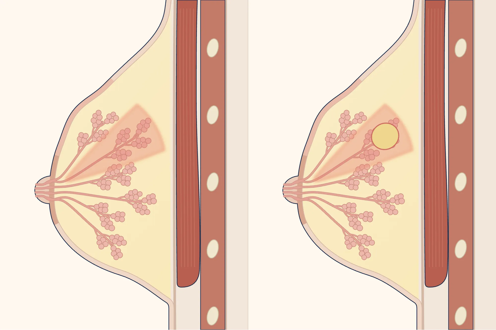
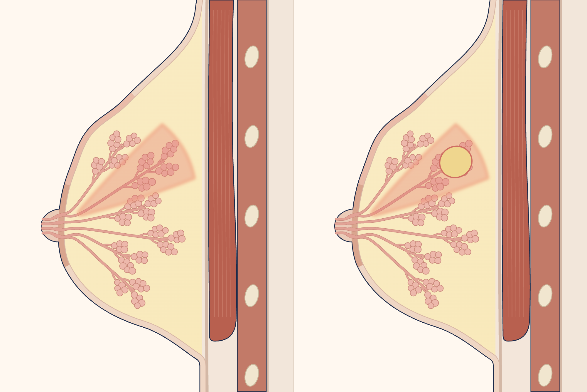

What is mastitis?
Mastitis is inflammation of the breast[1]. It occurs most often during breastfeeding, especially in the first weeks after birth, but it can occur at any stage of breastfeeding[2, 13]. Mastitis during breastfeeding is a spectrum ranging from narrowing of the milk ducts to inflammation, bacterial infection and a collection of pus (abscess)[1]. About 3 to 11 per cent of women who have mastitis develop an abscess[1].
What are the symptoms of mastitis?
Mastitis usually starts quickly and in one breast[13]. Part of the breast becomes swollen, warm and painful to the touch; it may become red, but redness is difficult to see on brown or dark skin[13]. There may also be a wedge-shaped area of firmness in the breast, a burning pain and white or blood-streaked discharge from the nipple[13].
Flu-like symptoms such as fever, chills and shivering, tiredness and body aches may accompany it[13]; these can also occur because of inflammation alone, without infection[1]. Fever is not always present[2].
Fullness caused by milk coming in, on the other hand, usually causes pain, firmness and swelling in both breasts at once on days 3–5 after birth; the Academy of Breastfeeding Medicine (ABM) considers this separate from mastitis[1]. Pain and redness seen in the morning after a long sleep are also usually not an infection[1].
The first 6 weeks after birth are a high-risk period for sepsis (a severe, potentially life-threatening response to infection), and sepsis can occur without a high fever[12, 16]. A rash that does not fade when pressed, a very low body temperature or passing no urine for 18 hours can also be signs of sepsis: if these occur or sepsis is suspected, call 112 or go to the nearest emergency department; do not drive yourself to the emergency department[12, 16].
What causes mastitis?
The cause of mastitis during breastfeeding is debated: the World Health Organization (WHO) and the Cochrane review consider milk not being adequately removed from the breast an important cause[2, 3]; ABM states that this has not been proven, that producing too much milk and expressing to “empty” the breast increase inflammation, and that deep massage also increases the risk of an abscess[1]. Mastitis is not contagious; there is no evidence that it is caused by poor hygiene or a particular food[1].
An abscess can develop from mastitis that is not treated promptly; early treatment of mastitis may reduce this possibility[14]. Having had mastitis recently or an abscess before also increases the likelihood of an abscess[14].
What helps with mastitis?
Mastitis during breastfeeding often clears with measures taken at home[1, 13]. Continuing to breastfeed may help recovery[2, 14]; milk from the breast with mastitis is safe for the baby[1]. According to the WHO, breastfeeding is usually safe even if the milk looks as though it contains pus, while stopping breastfeeding may make the condition worse[2]. The following are recommended:
- Breastfeed whenever your baby wants and for as long as your baby wants[13].
- If breastfeeding is very painful or your baby will not take that breast, express enough milk by hand for your baby; do not express more milk than your baby needs[1, 13].
- Ask a midwife or breastfeeding counsellor for help with getting your baby to latch on correctly[13].
- Do not massage the breast deeply or press on it; use a light touch[1, 13].
- Do not apply oil, cream or salt water to the breast[1, 13].
- Paracetamol or ibuprofen can be used for pain and fever; do not take aspirin as a painkiller[1, 13].
- Rest; wear a comfortable, supportive bra and avoid tight clothing until you recover[13].
Sources differ on applying heat or cold: the United Kingdom National Health Service (NHS) recommends a cold compress for 10 minutes every hour[13]; the Academy of Breastfeeding Medicine (ABM) recommends ice and states that heat may increase symptoms but may provide relief for some women[1]; the WHO, on the other hand, recommends a warm compress to ease pain and help the milk flow[2]. How often milk should be removed is also debated: the WHO considers effective milk removal the foundation of treatment and recommends frequent breastfeeding[2]; ABM, on the other hand, says that excessive breastfeeding and expressing may increase inflammation[1].
If the area around the nipple is so swollen that milk cannot come out, ABM recommends feeding the baby from the other breast until the swelling goes down[1].
How is mastitis treated?
Antibiotics are not needed for every case of mastitis: the Academy of Breastfeeding Medicine (ABM) reserves antibiotics for bacterial mastitis because unnecessary use disrupts the balance of bacteria in the breast and makes it easier for resistant germs to develop[1]. According to the WHO, antibiotics are given if symptoms are severe from the outset or do not improve within 12–24 hours despite better milk removal[2].
Take the antibiotic for as long as your doctor tells you to: according to the World Health Organization (WHO), delayed or incomplete treatment increases the likelihood of an abscess and recurrence[2]. While breastfeeding, a very small amount of the antibiotic may pass into the milk; this does not usually pose a risk to the baby, but it may make the baby unsettled[1, 13]. If there is no improvement two days after starting the antibiotic, a test to grow germs from the milk (culture) may be requested[1, 2].
If mastitis does not go away despite treatment or recurs in the same place, imaging and, if necessary, a biopsy are used to check for an underlying lump, granulomatous mastitis or, rarely, cancer[1, 4].
How is a breast abscess drained, and is surgery needed?
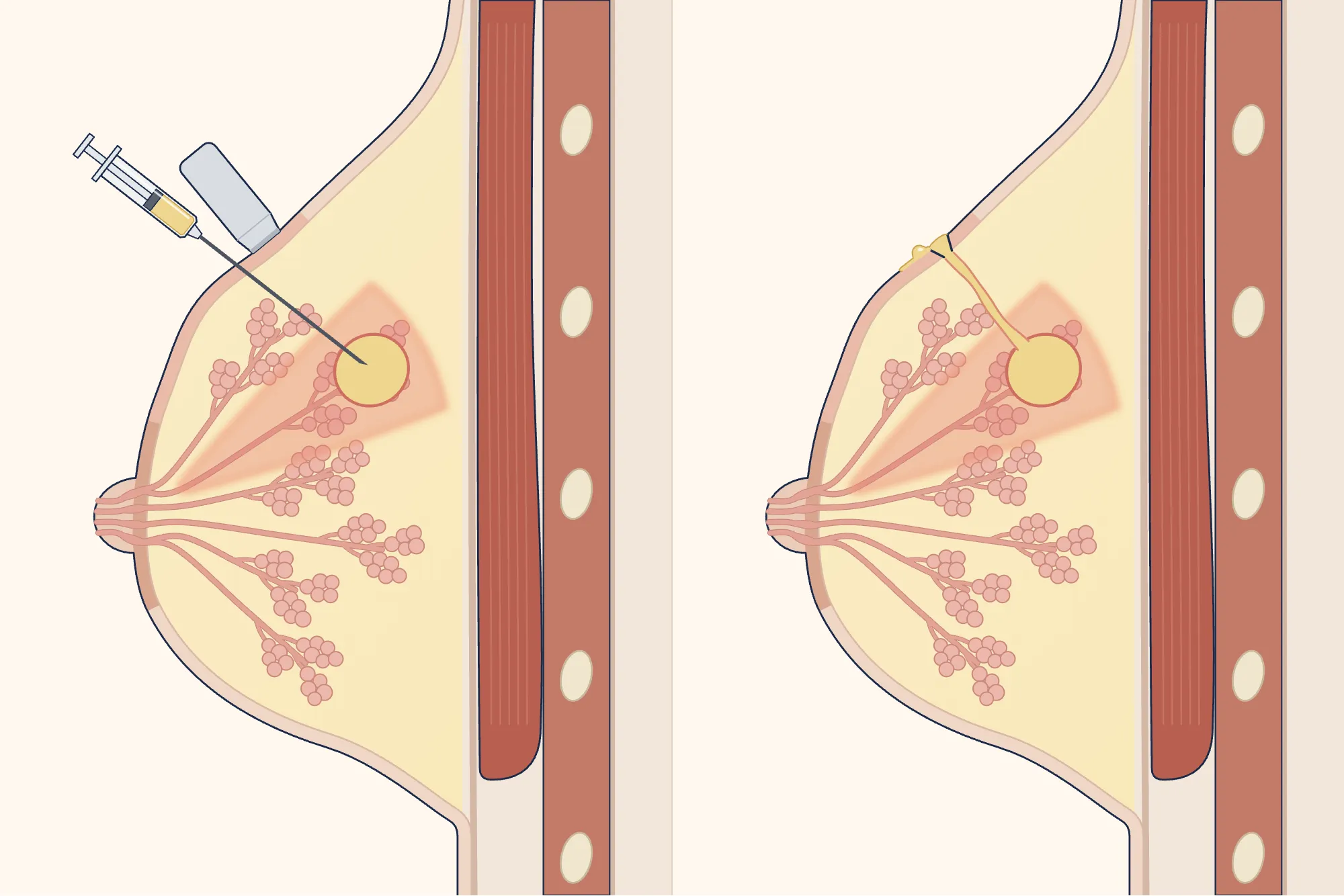
The pus must be drained; antibiotics alone are not expected to help without drainage[1, 2]. The pus is drawn out with a needle, usually with ultrasound guidance, or drained through a small cut in the skin[2, 14]. Needle drainage is performed under local anaesthetic, usually as an outpatient, and may need to be repeated several times; drainage through a cut can be performed under local or general anaesthetic; most people go home the same day[2, 3, 14].
Enlarge
Swipe the drawing right or left to see the details. You can use the arrow keys on your keyboard.
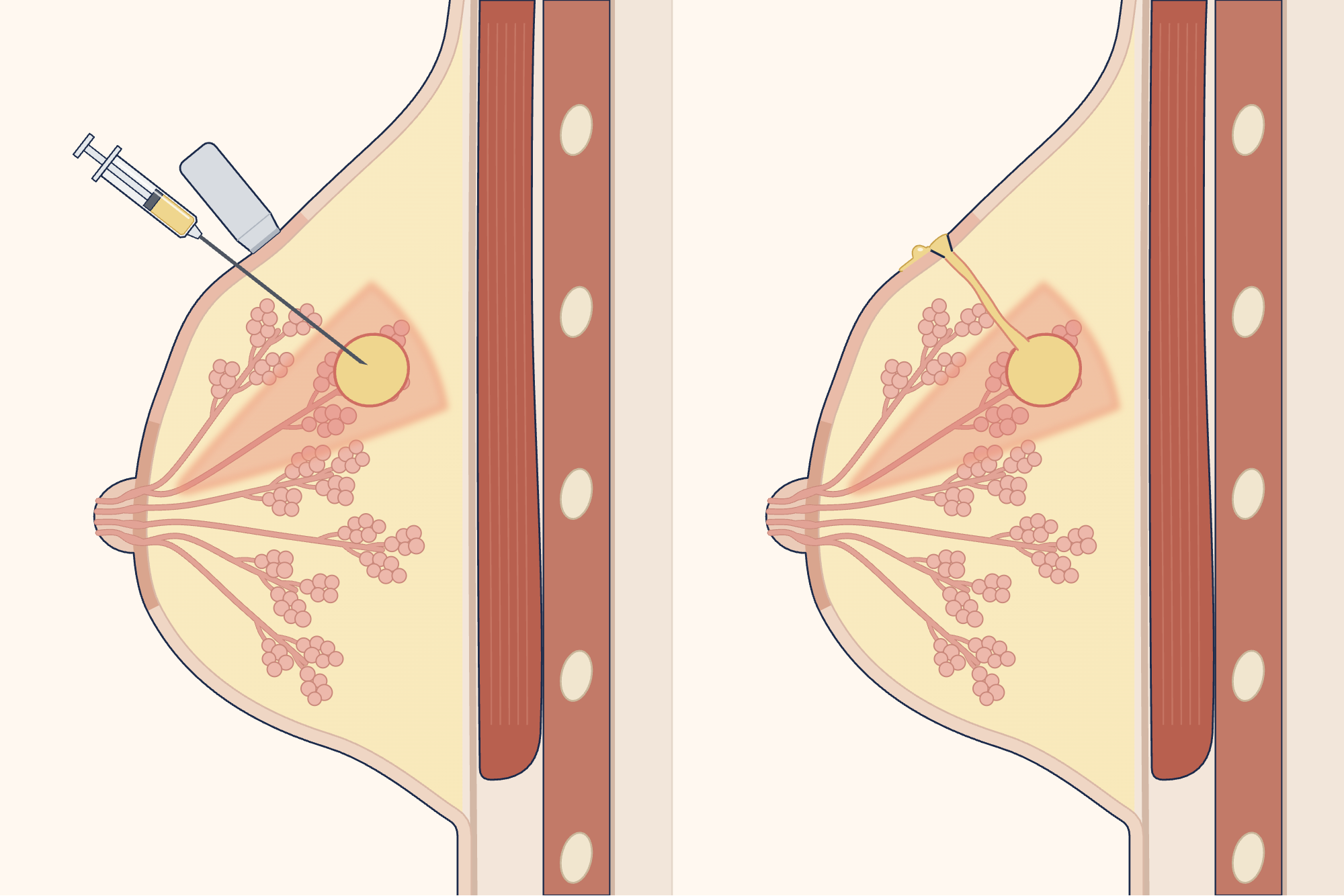
- 1 Pocket of pus (abscess)
- 2 Ultrasound probe
- 3 Needle
- 4 Withdrawn pus
- 5 Small cut in the skin
- 6 Pus flowing out
- 7 Nipple
Description of the drawing
The pocket of pus in the breast (abscess) must be drained. Left: while the ultrasound probe shows the pocket, a needle is advanced through the skin into it and the pus is drawn into a syringe. Right: pus flows out through a small incision made in the skin over the pocket.
Results may vary from person to person with any surgical or interventional procedure. You are advised to discuss the procedure in detail with your doctor beforehand.
Illustrative drawing, not to scale. Source: Cancer Research UK / Wikimedia Commons, CC BY-SA 4.0. English text and pointer lines were removed; the breast outline and nipple position from the source were retained, with only the lower end redrawn to complete the fold beneath the breast; the skin, nipple and surrounding ring, milk glands, ducts, chest muscle and ribs were redrawn, and the fatty tissue was coloured; the inflamed wedge, the reddened skin over it and the pocket of pus were added; an ultrasound probe, needle and syringe were added to the left panel, and a small cut, channel and outflowing pus to the right panel; numbered labels were added. This adaptation is also licensed under CC BY-SA 4.0.
An abscess is a collection of pus in the breast caused by infection; it is not usually serious, but requires treatment in hospital[14]. It can cause a painful lump or swelling in the breast, warmth or redness, fever and a general feeling of being unwell[14]; the fever may fall, or fall and rise again[1]. If a painful swelling in the breast appears and continues to grow during mastitis, see a doctor the same day[1, 14].
If an abscess is suspected, ultrasound is the first examination performed[4, 14]. If you are over 30 or cancer is suspected, a mammogram, which is also safe while breastfeeding, may be performed because an abscess can resemble inflammatory breast cancer[4].
A 2015 Cochrane review found insufficient evidence to determine which method was better; treatment failed more often with needle drainage (low-quality evidence), while breastfeeding was continued more often in one study[3]. The American College of Radiology (ACR) considers needle drainage the first option in most cases because healing rates are similar and fistulas and interruptions to breastfeeding are less common[4]. Drainage through a cut may be needed for large or multiple abscesses[3, 5].
Continuing to breastfeed from the affected breast after the procedure is recommended[1, 2]; the baby's mouth should not touch the wound or pus[3]. Milk leaking from the wound (milk fistula) occurs in fewer than 2 in 100 women after drainage with a needle or tube (drain)[1]. The abscess is expected to heal completely within a few days to a few weeks[14]; because a small area of firmness may be felt in the breast for weeks, healing is monitored with examination and imaging[1].
What causes mastitis when you are not breastfeeding?
Mastitis can also occur in women who are not breastfeeding and in men[13]. Smoking, damage to the nipple (for example, from piercing or eczema), a breast implant, a condition that weakens the immune system such as diabetes, and removing hair around the nipple by shaving or plucking can cause it[13].
The two main types of mastitis outside breastfeeding are inflammation of the milk ducts around the nipple (periductal mastitis), which is seen mostly in people who smoke, and granulomatous mastitis[5, 6]. Both cause lumps or abscesses, last approximately 9–12 months and recur frequently[5]. Which treatment is more suitable for periductal mastitis is debated; the options are stopping smoking, antibiotics and drainage of the pus, while surgery is reserved for cases that do not resolve[6].
Redness and swelling in the breast when you are not breastfeeding can be confused with inflammatory breast cancer: many women with this type of cancer are initially misdiagnosed with mastitis[7]. For this reason, ultrasound is performed, as well as a mammogram if cancer is suspected; the diagnosis is confirmed with a tissue sample taken using a thick needle (biopsy)[5, 7].
What is granulomatous mastitis?
Granulomatous mastitis of unknown cause (idiopathic granulomatous mastitis) is a long-lasting, benign inflammation of the breast[8, 11]. It is rare, but is reported more often in some regions, such as Türkiye[8, 10, 11]. It begins with a lump in the breast that may or may not be painful; the lump may grow rapidly and become red, and as it progresses, abscesses and openings in the skin that discharge fluid may develop; there is usually no fever[8]. Because it can resemble cancer, it is diagnosed by biopsy[9, 11]; sources differ on whether tests for tuberculosis (TB) are needed in every patient[8, 9, 11].
In up to half of patients, the condition resolves without treatment within 2–24 months[10]. According to the Turkish consensus, the first option is close monitoring for small-scale disease and during pregnancy or breastfeeding, and cortisone-type medicines for widespread disease with signs such as skin wounds; abscesses are drained[10, 11]. The role of surgery is debated: the international consensus recommends wide surgical removal for selected complex lesions[8]; the Turkish consensus, however, recommends that surgery should not be the first treatment for disease that is not widespread and states that the condition recurs in 23–50% of cases after limited removal[10].
Which department treats mastitis?
The general surgery department treats inflammation and abscesses in the breast; this department or radiology drains the abscess, and ultrasound and mammograms are also performed in radiology. If you are breastfeeding, your family doctor or workplace doctor can carry out the initial examination; if they consider it necessary, they may refer you to general surgery.
Sources
19
- Mitchell KB, Johnson HM, Rodríguez JM, Eglash A, Scherzinger C, Widmer K, et al. Academy of Breastfeeding Medicine Clinical Protocol #36: The Mastitis Spectrum, Revised 2022. Breastfeed Med. 2022;17(5):360–376. doi: 10.1089/bfm.2022.29207.kbm. PubMed: 35576513.
- World Health Organization (WHO). Mastitis: causes and management. WHO/FCH/CAH/00.13. Geneva: WHO; 2000. Text: iris.who.int.
- Irusen H, Rohwer AC, Steyn DW, Young T. Treatments for breast abscesses in breastfeeding women. Cochrane Database Syst Rev. 2015;(8):CD010490. doi: 10.1002/14651858.CD010490.pub2. PubMed: 26279276.
- Expert Panel on Breast Imaging: Dogan BE, Salkowski LR, Weinstein SP, et al. ACR Appropriateness Criteria: Breast Imaging During Lactation. J Am Coll Radiol. 2026;23(8):1768–1785. doi: 10.1016/j.jacr.2026.03.002. PubMed: 41885663.
- Zhou F, Shang XC, Tian XS, Yu ZG; Chinese Society of Breast Surgery. Clinical practice guidelines for diagnosis and treatment of patients with non-puerperal mastitis: Chinese Society of Breast Surgery (CSBrS) practice guideline 2021. Chin Med J (Engl). 2021;134(15):1765–1767. doi: 10.1097/cm9.0000000000001532. PubMed: 34039865.
- Mitchell KB, Johnson HM, Eglash A; Academy of Breastfeeding Medicine. ABM Clinical Protocol #30: Breast Masses, Breast Complaints, and Diagnostic Breast Imaging in the Lactating Woman. Breastfeed Med. 2019;14(4):208–214. doi: 10.1089/bfm.2019.29124.kjm. PubMed: 30892931.
- Dawood S, Merajver SD, Viens P, Vermeulen PB, Swain SM, Buchholz TA, et al. International expert panel on inflammatory breast cancer: consensus statement for standardized diagnosis and treatment. Ann Oncol. 2011;22(3):515–523. doi: 10.1093/annonc/mdq345. PubMed: 20603440.
- Yuan QQ, Xiao SY, Farouk O, Du YT, Sheybani F, Tan QT, et al. Management of granulomatous lobular mastitis: an international multidisciplinary consensus (2021 edition). Mil Med Res. 2022;9(1):20. doi: 10.1186/s40779-022-00380-5. PubMed: 35473758.
- Emiroglu M, Akcan A, Velidedeoglu M, Girgin S, Aytac O, Canturk NZ, et al. Diagnosis, Approach, and Clinical Classification of Idiopathic Granulomatous Mastitis: Consensus Report. Breast Care (Basel). 2024;19(5):243–251. doi: 10.1159/000541482. PubMed: 39439860.
- Emiroglu M, Cetin K, Yilmaz KB, Velidedeoglu M, Girgin S, Akcan A, et al. Idiopathic Granulomatous Mastitis: A Consensus Report on Treatment and Follow-Up Approaches Based on the Turkish Clinical Classification. Breast Care (Basel). 2025;20(4):248–255. doi: 10.1159/000544967. PubMed: 40860773.
- Emiroğlu M, Yılmaz KB, Çetin K, Velidedeoğlu M, Esendağlı G, Kayadibi Y, et al. Idiopathic granulomatous mastitis: A narrative review based on the Turkish consensus classification. Turk J Surg. 2026;42(1):1–12. doi: 10.47717/turkjsurg.2026.2025-8-40. PubMed: 41766585.
- National Institute for Health and Care Excellence (NICE). Suspected sepsis in pregnant or recently pregnant people: recognition, diagnosis and early management. NICE guideline NG255, 19 November 2025. Text: www.nice.org.uk.
- NHS (United Kingdom National Health Service). Mastitis: patient information page. Last reviewed: 17 March 2023. Text: www.nhs.uk.
- NHS (United Kingdom National Health Service). Breast abscess: patient information page. Last reviewed: 14 June 2023. Text: www.nhs.uk.
- NHS (United Kingdom National Health Service). Breast pain: patient information page. Last reviewed: 3 May 2023. Text: www.nhs.uk.
- NHS (United Kingdom National Health Service). Sepsis: patient information page. Last reviewed: 14 May 2026. Text: www.nhs.uk.
- healthdirect Australia (health information service supported by the Australian government). Mastitis: patient information page. Last reviewed: April 2024. Text: www.healthdirect.gov.au.
- National Cancer Institute (NCI). What Is Breast Cancer?: breast anatomy section of the official patient information. Last updated: 2 December 2025. Full page accessed: 4 October 2026. Text: www.cancer.gov.
- National Cancer Institute (NCI), SEER Training Modules. Anatomy: Anatomy of the Female Breast. Last updated: 10 January 2025. Full page accessed: 4 October 2026. Text: training.seer.cancer.gov.
Medical review of the Turkish original: Op. Dr. Ömer Seyfullah Hasıripi, . This page is a translation checked by AI reviewers; the Turkish version prevails.
This page is for general information; it does not replace an examination. See a doctor about your symptoms.