Illustration sources
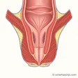
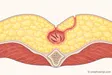
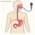
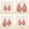
Illustrative drawings prepared for this site, not to scale: Piles, Pilonidal sinus, Anal tear, Gastroscopy, Groin hernia, Umbilical hernia, Abdominal wall hernias, Gallstones, Goitre and thyroid nodule, Breast enlargement in men.
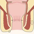
Anal abscess: Illustrative drawing, not to scale. Source: Armin Kübelbeck, McortNGHH and Jmarchn, CC BY-SA 4.0. Adapted from Jmarchn’s “Abscess diag 02-es” drawing (CC BY-SA 3.0), which is based on McortNGHH’s “Abscess diag 02” (CC BY-SA 4.0) and Armin Kübelbeck’s “Piles diffdiag 01” (CC BY 3.0). The text, arrows and abscess markings in the source were removed; the right half was mirrored to create a base with identical sides, the side outline of the body was redrawn, the colours were changed, the band between the two muscles was widened, and anal glands opening into the canal at the level of the dentate line and four abscess sites were added; numbered labels were added. This adaptation is also licensed under CC BY-SA 4.0.
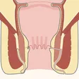
Anal fistula: Illustrative drawing, not to scale. Source: Armin Kübelbeck, McortNGHH and Jmarchn, CC BY-SA 4.0. Adapted from Jmarchn’s “Abscess diag 02-es” drawing (CC BY-SA 3.0), which is based on McortNGHH’s “Abscess diag 02” (CC BY-SA 4.0) and Armin Kübelbeck’s “Piles diffdiag 01” (CC BY 3.0). The text, arrows and abscess markings in the source were removed; the right half was mirrored to create a base with identical sides, the side outline of the body was redrawn, the colours were changed, the band between the two muscles was widened, and anal glands opening into the canal at the level of the dentate line, the spread of an abscess on one side and a fistula tunnel on the other were added; numbered labels were added. This adaptation is also licensed under CC BY-SA 4.0.
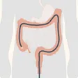
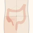
Colonoscopy: Illustrative drawing, not to scale. Source: Cancer Research UK / Wikimedia Commons, CC BY-SA 4.0. The magnified inset, tumour, wire loop, handle of the instrument and English text were removed, the small intestine and stomach were faded, and the image was cropped to a square; the large bowel was coloured in a single colour, the anal canal was widened slightly, the colonoscope tube was redrawn from the anus to the beginning of the large bowel, and a light was added to its tip; numbered labels were added. This adaptation is also licensed under CC BY-SA 4.0.
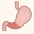
Gastritis: Illustrative drawing, not to scale. Source: Cancer Research UK / Wikimedia Commons, CC BY-SA 4.0. The English text and pointer lines were removed and the colours were changed; the oesophagus and duodenum were shown cut off with a straight line, and the portion beyond the descending part of the duodenum was removed; muscle and inner lining layers were added to the stomach wall, folds to the inner surface, a ring of muscle at the stomach outlet, and inflamed lining and a boundary line to the lower part of the stomach; numbered labels were added. This adaptation is also licensed under CC BY-SA 4.0.
Appendicitis: Illustrative drawing, not to scale. Source: Cancer Research UK / Wikimedia Commons, CC BY-SA 4.0. The English text and organs other than the large bowel were removed, the large bowel was faded and the caecum made more prominent, and the outer outline of the rectum and the bend where the large bowel joins the rectum were redrawn as a smooth tube; a body silhouette, faint arcs representing the ribs and hip bones, a belly button, dashed lines dividing the abdomen into four sections, colouring in the lower right section, the final part of the small intestine and a new appendix were added; numbered labels were added. This adaptation is also licensed under CC BY-SA 4.0.
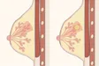
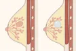
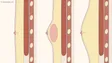
Breast inflammation: Illustrative drawing, not to scale. Source: Cancer Research UK / Wikimedia Commons, CC BY-SA 4.0. The English text and pointer lines were removed; the breast outline and nipple position from the source were retained, with only the lower end of the outline redrawn to complete the fold beneath the breast; the skin, nipple and the ring around it, milk glands, ducts, chest muscle and ribs were redrawn, and the fatty tissue was coloured; an inflamed wedge, its slight swelling (the clusters of glands within it were spaced a little further apart and the skin above it raised slightly), the reddened skin over it and a pocket of pus were added; numbered labels were added. This adaptation is also licensed under CC BY-SA 4.0.
Breast lump: Illustrative drawing, not to scale. Source: Cancer Research UK / Wikimedia Commons, CC BY-SA 4.0. The English text and pointer lines were removed; the breast outline and nipple position from the source were retained, with only the lower end of the outline redrawn to complete the fold beneath the breast; the skin, nipple and the ring around it, milk glands, ducts, chest muscle and ribs were redrawn, and the fatty tissue was coloured; a fibroadenoma and cyst were added, and the surrounding glands were moved aside; numbered labels were added. This adaptation is also licensed under CC BY-SA 4.0.
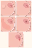
Breast cancer symptoms and screening: Illustrative drawing, not to scale. Source: Hariadhi / Wikimedia Commons, CC BY-SA 4.0. The English text was removed; the panels were cropped slightly closer to the breast and arranged in two columns; the lump in the first panel, the indentation in the second, the nipple retraction in the fourth and the discharge in the fifth were redrawn; the dots in the third panel were redrawn as skin dimples; the background was changed to cream; numbered labels were added. This adaptation is also licensed under CC BY-SA 4.0.
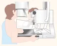
Breast imaging and biopsy: Illustrative drawing, not to scale. Source: Cancer Research UK / Wikimedia Commons, CC BY-SA 4.0. The “Cancer Research UK” text in the corner of the source was removed; the upper surface of the breast was flattened slightly to show that the plate touches it, its lower edge was flattened a little, and a shadow was added where it rests on the platform; the background was changed to cream and the blank edges were cropped; numbered labels were added. This adaptation is also licensed under CC BY-SA 4.0.