What is a pilonidal sinus?
A pilonidal sinus (also known medically as pilonidal disease) is a relatively common and benign condition found in the cleft between the buttocks near the tailbone[3]. The Dutch guideline defines it as a disease caused by hairs penetrating beneath the skin; the disease leads to recurrent inflammation or an abscess, often with a discharge of pus or blood[1].
One or more small pits can be seen in the middle of the cleft between the buttocks[1]; tracts extend from these pits beneath the skin, often containing hair and skin debris[6]. Some patients may also have openings in the skin of the buttocks away from the midline[1, 3]. Most people do not notice a pilonidal sinus until it becomes inflamed and causes symptoms[4].
Enlarge
Swipe the drawing right or left to see the details. You can use the arrow keys on your keyboard.
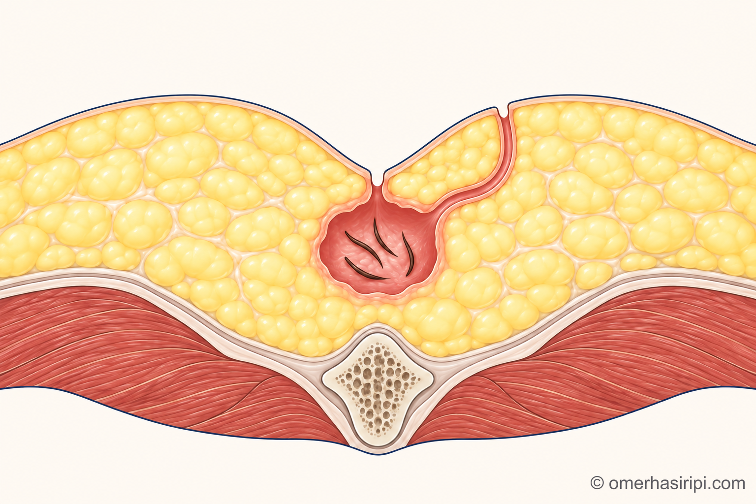
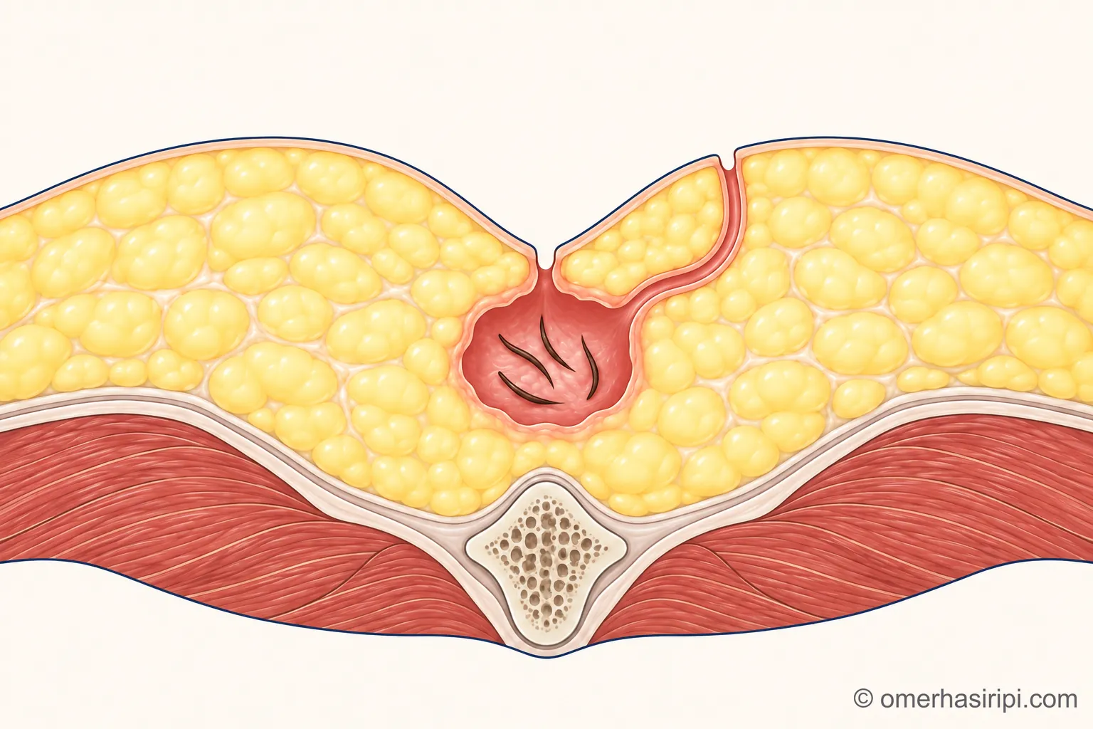
- 1 Skin
- 2 Pit in the midline
- 3 Cavity filled with hair
- 4 Tract leading to the side
- 5 Fat tissue beneath the skin
- 6 Tailbone
Description of the drawing
Cross-sectional drawing: the pit in the midline opens into a cavity beneath the skin containing hairs; a narrow tract leading from the cavity may end at a second opening in the skin at the side.
Illustrative drawing, not to scale. Prepared for this site.
What causes it and who gets it?
It is now generally accepted that the disease is not present at birth but develops later, and that hairs entering through the skin in the cleft between the buttocks play a role[1]. The cavity beneath the skin usually contains short pieces of hair with cut ends and no roots; these hairs resemble scalp hair more than body hair[1]. According to the German guideline, cut pieces of hair can be found on the shoulders, back and lower back, that is, on the back of the body, within minutes of a haircut[2]. Three factors stand out in the development of the disease: loose hairs falling into the cleft between the buttocks, friction, and the ease with which the skin in this area is damaged[1].
The disease occurs mostly in young people and young adults; the age at which it first appears is increasingly shifting towards 15–25 years, and among adults it is about four times more common in men[2]. Pilonidal sinus is rare before puberty[2]. It is said to be more common in people with a lot of body hair, but it also occurs in people with little body hair[1]. A family history of pilonidal sinus, obesity (excess weight) and smoking are also listed among the factors that make the disease more likely[1, 8]. Sitting for long periods is also mentioned, but there is almost no research on this[1]. The Dutch guideline considers these factors alongside expert opinion without systematically evaluating the research[1].
A deep cleft between the buttocks is also mentioned as a factor that makes the disease more likely[1]; however, based on a more recent and larger measurement study, the German guideline states that the depth of the cleft is not a risk factor. According to older studies, the thickness of the fat tissue over the tailbone appears to be a risk factor[2].
Poor hygiene and heavy sweating have also been suggested to make people more prone to the disease; however, people with a pilonidal sinus have also been found to sweat less than similar people[1]. The German guideline states that there is no evidence that hygiene has a direct role in the development, flare-up or return of the disease; regular removal of sharp hair fragments from between the buttocks by showering or bathing, however, appears to be helpful[2].
How can you recognise it and what are the symptoms?
Symptoms include pain, swelling, bloody discharge and itching[1]. The German guideline distinguishes three forms of the disease: disease that causes no symptoms, an abscess that develops suddenly, and long-standing (chronic) disease; the most common form is long-standing disease with intermittent discharge[2].
When an abscess develops suddenly, the area around the tailbone becomes red, tender to the touch and swollen; fluid can be felt within the swelling, and sometimes there is also a fever[3]. An abscess can develop suddenly and make sitting difficult[4]. If inflammation happens repeatedly, pain and discomfort may persist; there is intermittent discharge of pus or blood[4]. The disease and its return may also lead to time away from work, school and sport[1].
Which department treats a pilonidal sinus?
A general surgery specialist assesses pilonidal sinus. You can also see your family doctor or occupational health doctor for an initial assessment. They can refer you to the relevant specialist when needed. If you have emergency symptoms, follow the advice on where to seek help in the “When is it an emergency?” section below.
How is it diagnosed?
The diagnosis is made by listening to your symptoms and examining you[1, 2, 3, 8]; blood tests or imaging are not routinely needed[2, 3, 8]. If the openings are close to the anus, an examination of the anus or imaging should be carried out to distinguish a pilonidal sinus from other anal conditions, such as an anal fistula[3, 8]. The assessment takes into account the number of openings, their position relative to the midline, their distance from the anus and any scars from previous surgery[3].
A pilonidal sinus can look very similar to another skin condition that causes repeated inflammation in the armpits and groin (hidradenitis suppurativa, commonly known in Turkish as “dog's breast” disease[10]); the two conditions can also occur together[1]. For this reason, the Dutch guideline recommends asking at the first examination whether there is recurrent inflammation in the armpits or groin, and involving a dermatologist in treatment if this condition is suspected[1]; the European Society of Coloproctology also recommends examining these areas if there is such a history[8].
Openings that cause no symptoms
If small openings seen by a doctor in the cleft between the buttocks cause no symptoms such as pain, itching or discharge, this is called a pilonidal sinus without symptoms[1]. The Dutch and German guidelines and the European Society of Coloproctology do not recommend surgery or preventive treatment in this situation[1, 2, 8]. According to the German guideline, a pilonidal sinus without symptoms does not go away on its own, can last a lifetime and may develop over time into a sudden abscess or long-standing disease; even so, preventive treatment is not needed[2]. The European Society of Coloproctology, unlike the German guideline, states that disease without symptoms may occasionally regress on its own[8]. The Dutch guideline states that these people may be advised to take the everyday measures described in the “Protecting yourself” section below instead of having surgery[1].
A sudden abscess
When an abscess develops suddenly, it needs to be opened with a small cut and adequately drained without delay[2, 3]. The German guideline states that published studies support making the cut beside the midline, and the European Society of Coloproctology also recommends drainage through a cut to the side[2, 8]; a large amount of tissue is not removed at this time[2]. Abscesses close to the surface can be drained after numbing the area; larger and deeper abscesses can be drained under a general anaesthetic. If needed, medicines to help you relax can also be added to the local anaesthetic[2]. This method of providing relaxation and a sleepy state together with medicines that reduce pain is called sedation and analgesia; the team assessing you will determine the appropriate method[13]. Afterwards, the wound needs regular dressings, and antibiotics are sometimes given[4]. Antibiotics may be considered for inflammation, but they do not cure the disease[8].
For small abscesses, the German guideline states that definitive treatment can also be carried out during the initial treatment[2]; the Italian consensus statement also considers cleaning out the abscess cavity or removing the diseased tissue at the first treatment a valid option for some patients[3]. If definitive treatment is to be carried out, it is usually planned after the inflammation and swelling have subsided (for example, after 10–14 days); according to the German guideline, this decision can also be made after the abscess wound has healed; definitive treatment carried out under emergency conditions may be associated with more frequent recurrence than planned treatment[2].
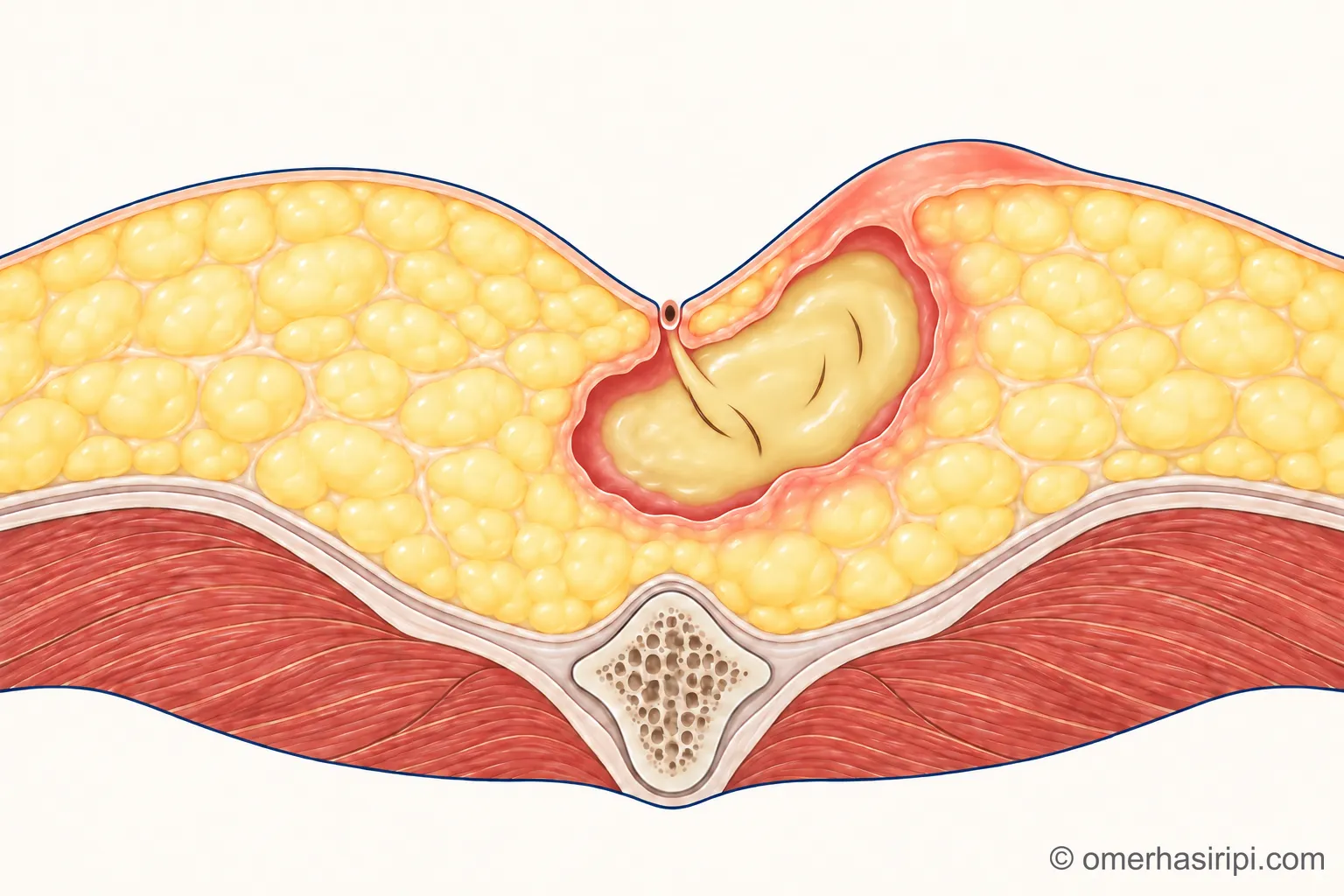
Enlarge
Swipe the drawing right or left to see the details. You can use the arrow keys on your keyboard.
- 1 Abscess (a collection of pus)
- 2 Red and swollen skin
- 3 Pit in the midline
Description of the drawing
A sudden abscess: the cavity beneath the skin fills with pus, and the skin above it becomes red, swollen and painful.
Illustrative drawing, not to scale. Prepared for this site.
What happens after drainage varies from person to person. According to the Italian consensus statement, the disease returns in a substantial proportion of patients who have drainage alone; in two comparative studies cited by the statement, the rates in the groups who had drainage alone were over 40% (with follow-up of up to 65 months) and 55%[3]. In two more recent studies cited by the German guideline, however, approximately two thirds of patients whose abscesses were drained did not require further surgery in the long term (an average of approximately five years in one study)[2]. These rates cannot be directly compared because the studies looked at different outcomes (recurrence of the disease or the need for further surgery) and different follow-up periods. Views also differ: according to the German guideline's recommendation, either waiting or definitive treatment can be chosen after the abscess wound has healed, meaning that definitive treatment is not mandatory for everyone; the guideline also states that healing on its own after adequate drainage is not uncommon[2]; the European Society of Coloproctology, on the other hand, recommends definitive treatment after drainage[8]. The International Society of Laser Proctology recommends that SiLaC (Sinus Laser Closure), a method in which a thin laser tip is advanced into the pilonidal sinus tract to close it, should not be used during a sudden abscess[6].
Treatment options for long-standing disease
There is currently no method that meets every expectation: a method that is simple and painless, has a wound that heals quickly and has a low rate of recurrence has not yet been found[2]. Which operation is most suitable remains debated[3]. The German guideline calls for planned surgery to be recommended for long-standing disease[2]; the European Society of Coloproctology also recommends considering definitive surgical treatment for patients with symptoms[8]. The method is chosen according to the form and extent of the disease[2]; the Dutch guideline calls for the advantages and disadvantages of the methods to be discussed with the patient and the decision made together[1].
Minor procedures for limited disease
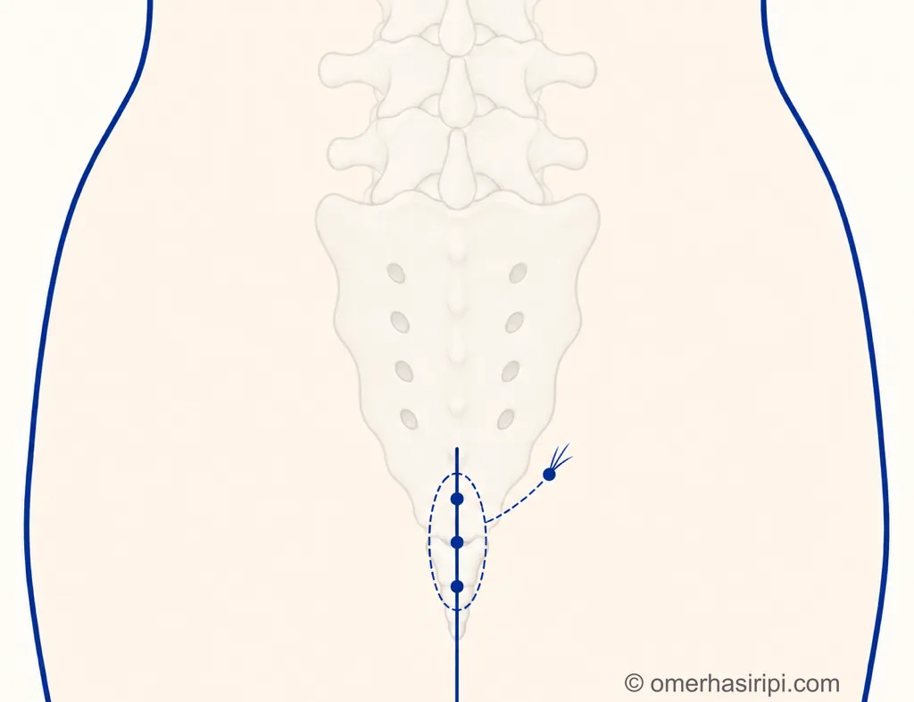
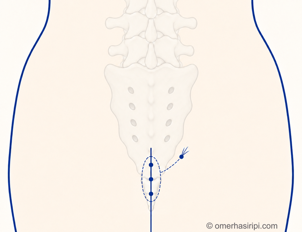
If the disease is limited and simple (for example, if there has been no previous surgery and there is one opening or a few openings in the midline), the Italian consensus statement considers minor procedures the first choice[3]; the Dutch guideline also recommends considering them as the first option[1]. The German guideline recommends offering a method called pit picking for limited disease; this method is particularly suitable for people who have not had surgery before and whose disease is less extensive[2]. In pit picking, the midline openings are removed with a margin of only about 1 millimetre of surrounding skin; the cavity beneath them is cleaned out, if necessary through a small cut made beside the midline[2]. The European Society of Coloproctology also states that minor procedures can be performed for limited disease and that pit picking can be considered; however, it considers the evidence underlying these recommendations to be very weak[8].
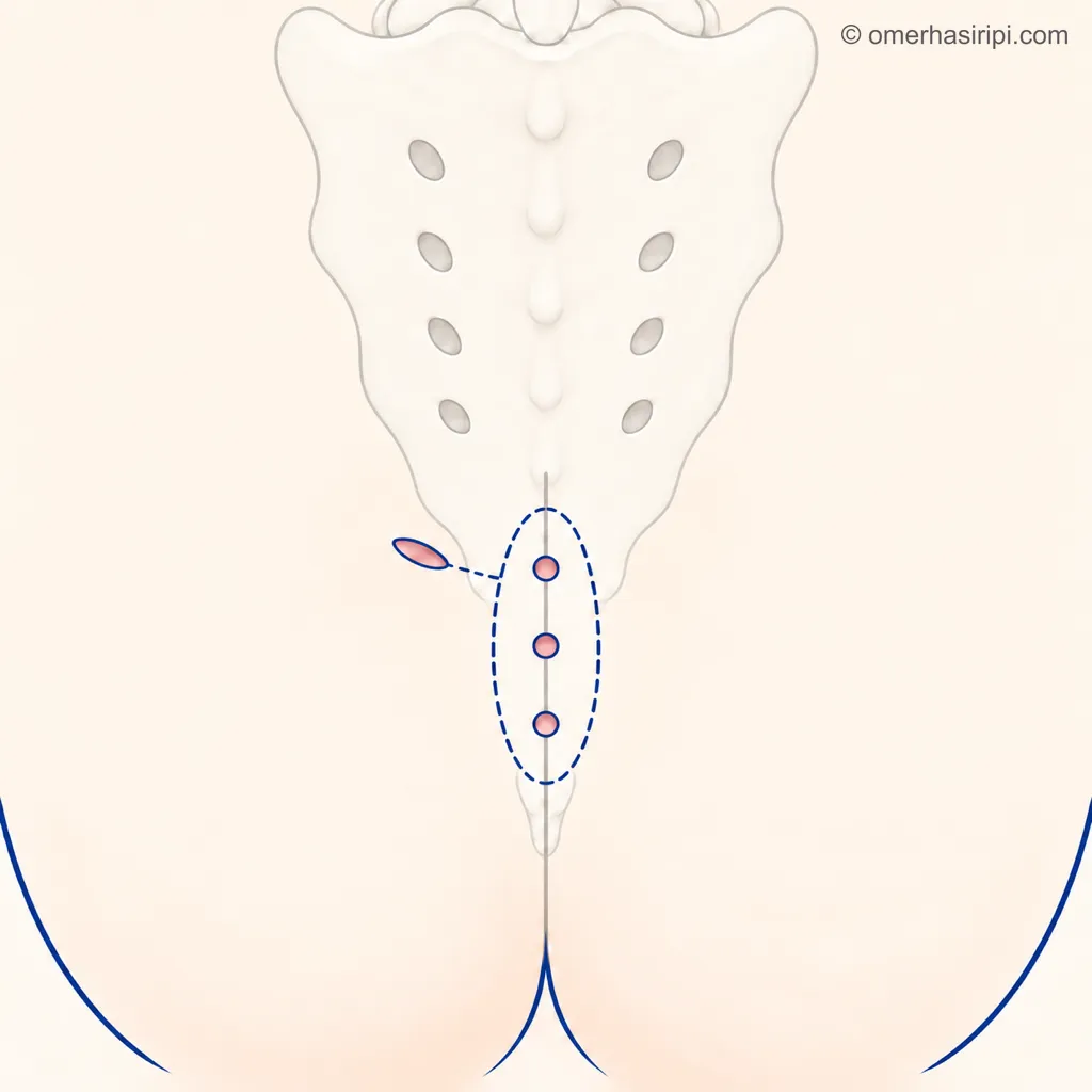
Enlarge
Swipe the drawing right or left to see the details. You can use the arrow keys on your keyboard.
- 1 Removal of the openings through very small cuts
- 2 Small opening through which the cavity is cleaned out
Description of the drawing
Minor procedure (for example, pit picking): the midline openings are removed through cuts a few millimetres across; the cavity beneath them is cleaned out through a small opening to the side if needed; no large wound is made. The dashed lines show the cavity beneath the skin and the connection extending to the opening at the side. The bones are drawn faintly only to show the location.
Results may vary from person to person with any surgical or interventional procedure. You are advised to discuss the procedure in detail with your doctor beforehand.
Illustrative drawing, not to scale. Prepared for this site.
These procedures can be performed under local anaesthetic without an overnight hospital stay[1, 2]; they place little burden on the patient and recovery is quick[8]. However, the German guideline states that the disease should be expected to return more often after these methods than after operations in which tissue is removed more extensively[2]; the Dutch guideline also says this appears to be the case compared with surgery in which tissue is removed and the wound is left open[1]: recurrence rates of approximately 8% to 16% have been reported after pit picking and similar methods over 1 to 10 years of follow-up; in some studies, rates reached 23–26% at three years[2]. The European Society of Coloproctology states that recurrence after minor procedures is not very different from that after larger operations, but depends greatly on the length of follow-up[8].
In the Gips method, the small openings are removed with a round-bladed skin cutter (biopsy punch), and the hair and debris beneath the skin are cleared out[3]. According to the Italian consensus statement, the strength of the method is that it can be repeated, even though the recurrence rate is high: in a large series, 85% of patients healed after one procedure, and a total of 95% healed after no more than two procedures[3]. The Dutch guideline also states that these methods cannot be strongly recommended because there is no robust evidence[1].
Laser, camera and phenol
New methods using a laser or a camera (endoscopic methods) are also considered minor procedures[3, 6, 8]. The European Society of Coloproctology states that both methods can be offered to patients, but considers the evidence for both to be weak or very weak[8]. The German guideline, however, states that the available evidence shows no advantage of either method over other minor procedures[2].
In reviews cited by the German guideline, treatment failed in approximately 8% of patients after camera treatment; in a study comparing camera treatment with “cleft lift” surgery, the disease returned in 24% of both groups over at least five years of follow-up[2]. The German guideline also reports that many other studies found recurrence rates of 0% to 11% for “cleft lift”; in three series in the guideline's table with follow-up of five years or longer (one involving only young people), the rate was 11% to 19%; the guideline also states that wound problems and recurrence should be expected in patients who have had several operations, whose wound has become chronic or who come to surgery with an open wound[2]. The same guideline also reports that wound healing problems after “cleft lift” are relatively common, with rates of 18% to 54% reported[2]. Studies report a healing rate of approximately 90% with laser treatment, but in one study 28% of patients required further surgery within a median of one year[2]. In studies cited by the Dutch guideline, the recurrence rate in patients who had laser treatment combined with removal of the openings ranged from 10–14% to 26%, and was approximately 37% in pooled five-year data[1]. Some positive views about these methods also come from those who perform them: the position paper of the International Society of Laser Proctology and an expert panel made up of surgeons who perform camera treatment[5, 6].
With the method in which a chemical called phenol is applied to the openings, the wound may heal more quickly; but its effect on recurrence of the disease compared with surgery in which the wound is left open is very uncertain[1]. The European Society of Coloproctology states that phenol can be offered, but that more than one application may be needed and that it has been withdrawn from use for this purpose in some countries[8]. In one analysis cited by the German guideline, phenol showed a recurrence rate similar to surgery and a shorter period of inability to work; another analysis found a recurrence rate of 40% five years after phenol treatment[2]. Because phenol is not approved for this use in Germany due to its toxic effects, the German guideline makes no recommendation, even though there is evidence in its favour[2].
Leaving the wound open
Operations in which the diseased tissue is removed at the midline are still the most commonly used methods today[2]. The method in which the tissue is removed and the wound is left open to heal on its own is safe, but the wound takes a long time to heal, especially if it is large: according to the German guideline, healing takes 1.5 to 3 months and an average of one month is spent away from work[2]. In studies, open wounds took 41 to 91 days to heal; frequent and painful dressing changes and close follow-up may be needed during this time[3]. Some open wounds may not heal for years[2].
The German guideline calls for tissue removal to be as limited as possible and recommends using methods that close the wound away from the midline (asymmetric methods) and flap operations instead of leaving it open[2]. The Italian consensus statement reserves leaving the wound open for complex cases[3]; the Dutch guideline also calls for caution with this method because of the long healing time[1]. The European Society of Coloproctology, however, states that leaving the wound open can be considered in selected patients[8]. Whether leaving the wound open reduces recurrence is debated[3].
Closing the wound and flap operations
The Dutch and German guidelines, the Italian consensus statement and the European Society of Coloproctology recommend that the wound should not be stitched directly on the midline after the diseased tissue is removed[1, 2, 3, 8]: when it is stitched on the midline, the wound opens or becomes inflamed more often and the disease returns more often[1, 2]. In studies cited by the German guideline, 14% to 74% of wounds stitched on the midline opened[2]. The Italian consensus statement considers closure away from the midline the gold standard if the wound is to be closed[3]; the European Society of Coloproctology also considers closure away from the midline the preferred method[8].
In methods that close the wound by moving it to the side of the cleft between the buttocks (flaps such as the Karydakis method, Bascom's “cleft lift” operation or a modified form of the Limberg flap), the scar is moved to the side of the midline and the upper part of the cleft is flattened. A cleft lift is an operation that makes the cleft between the buttocks shallower and moves the stitch line to the side of the cleft; to do this, depending on the method, nearby skin or skin together with the tissue beneath it is moved (a flap)[2, 8]. The German guideline recommends offering the Karydakis method and the modified form of the Limberg flap (the form that also moves the lower end of the wound line to the side of the cleft between the buttocks); it states that “cleft lift” surgery can also be considered[2]. The Dutch guideline recommends considering flap surgery for complex or recurrent disease and referring the patient to an experienced centre if there is no experience with it[1].
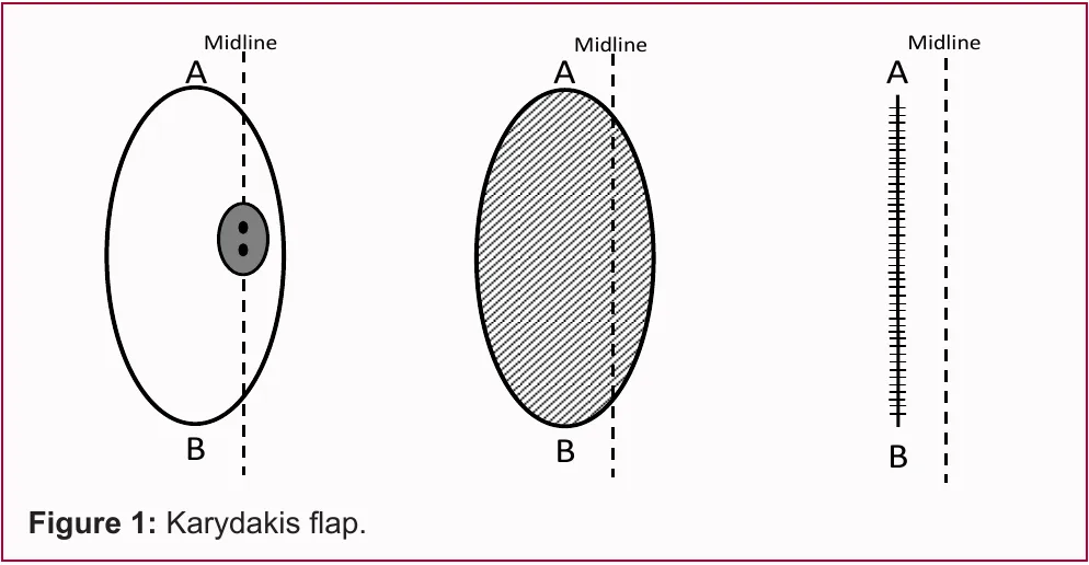
Enlarge
Swipe the drawing right or left to see the details. You can use the arrow keys on your keyboard.
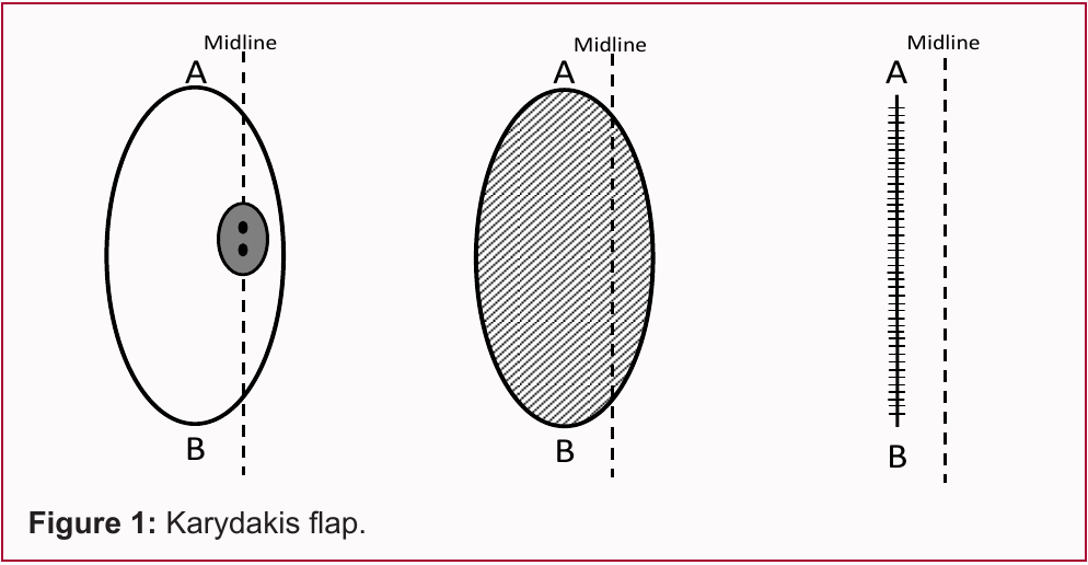
- 1 Dashed line: midline
- 2 Stitch line away from the midline
Description of the drawing
Off-midline closure in the Karydakis method is shown in a diagram of the lines on the skin surface. On the left is an asymmetrical oval plan encompassing the diseased area on the midline, in the centre is the area to be removed, and on the right is the suture line beside the midline. The dashed line is the midline. A and B in the three sections show the upper and lower ends of the oval area and the suture line. The English caption below means “Figure 1: Karydakis flap”. This figure does not show the bone or the depth of the operation; it is not an exact depiction of the final scar.[14, 15]
Results may vary from person to person with any surgical or interventional procedure. You are advised to discuss the procedure in detail with your doctor beforehand.
Illustrative drawing, not to scale. Source: Lim J and Shabbir J (2019), Figure 1, Creative Commons Attribution (version not specified in the source). The figure was extracted from the source PDF; the drawing elements were not changed. Numbered explanations in Turkish were added.
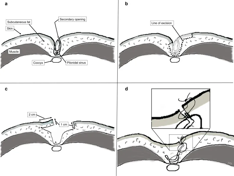
Enlarge
Swipe the drawing right or left to see the details. You can use the arrow keys on your keyboard.
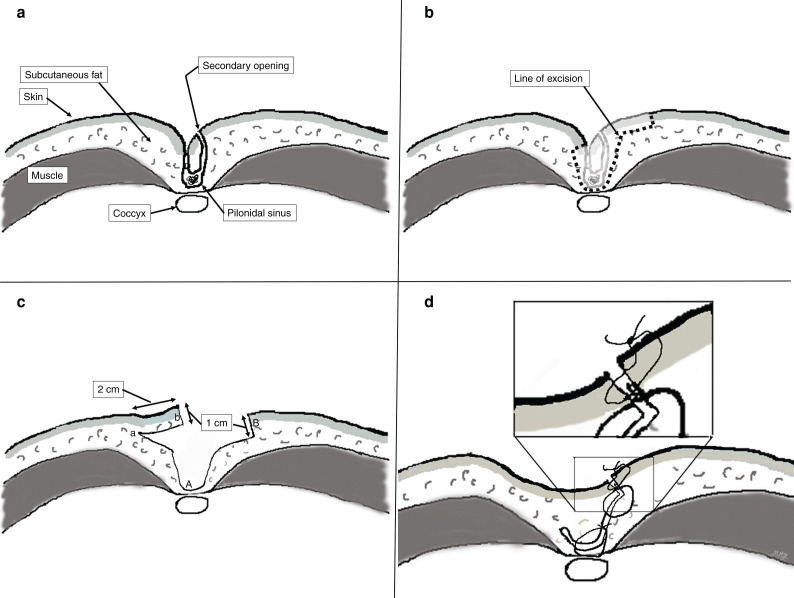
- 1 Cross-sectional view of the pilonidal sinus
- 2 Diseased area to be removed
- 3 Prepared flap of skin and tissue beneath the skin
- 4 Closure by advancing the flap
- 5 Upper layer of deep stitches securing the flap
- 6 Lower layer of deep stitches securing the flap
Description of the drawing
Cross-sectional diagram of the Karydakis method: a, the pilonidal sinus and surrounding tissues; b, the boundary of the diseased area to be removed; c, a flap of skin and tissue beneath the skin prepared from the side; d, closure of the wound by advancing and stitching the flap. In d, the two loops seen between the skin stitches and the tailbone represent two layers of deep stitches that hold the flap in place. The aim is to make the cleft between the buttocks shallower and move the closure line away from the midline. The English labels in the drawing are: Skin, skin; Subcutaneous fat, fat tissue beneath the skin; Muscle, muscle; Coccyx, tailbone; Secondary opening, secondary opening; Pilonidal sinus, pilonidal sinus; Line of excision, boundary of the area to be removed. The 2 cm and 1 cm in the drawing are the width and thickness of the flap prepared in the source study.[15]
Results may vary from person to person with any surgical or interventional procedure. You are advised to discuss the procedure in detail with your doctor beforehand.
Illustrative drawing, not to scale. Source: Hemmingsson et al. (2022), CC BY 4.0. The original source image was retained; numbered explanations in Turkish were added.
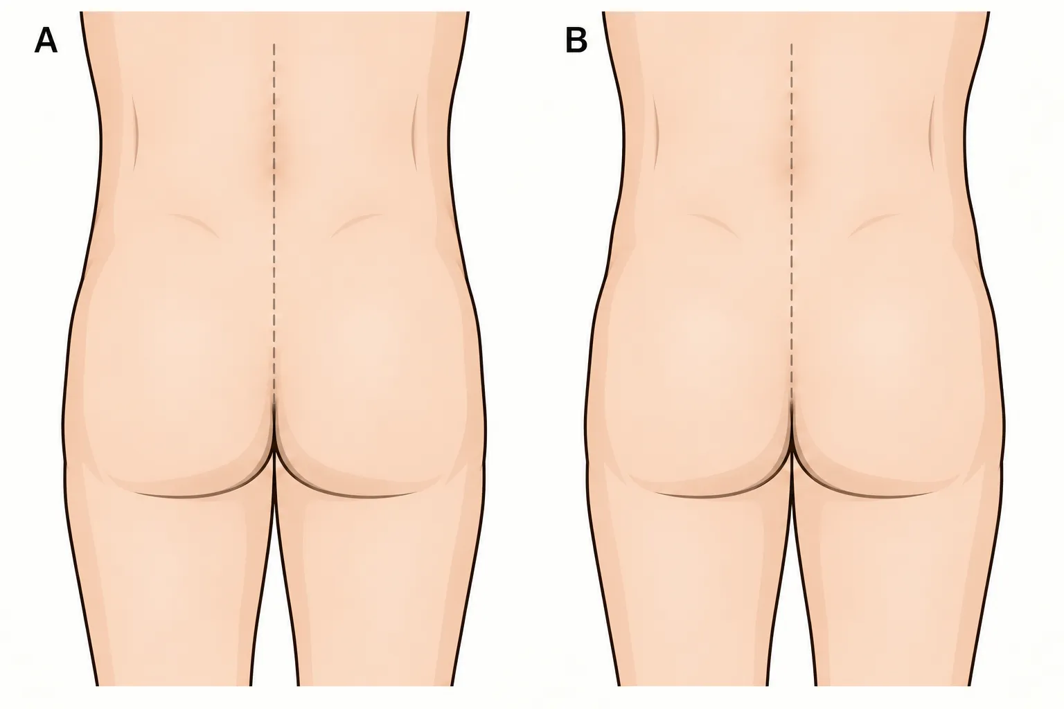
Enlarge
Swipe the drawing right or left to see the details. You can use the arrow keys on your keyboard.
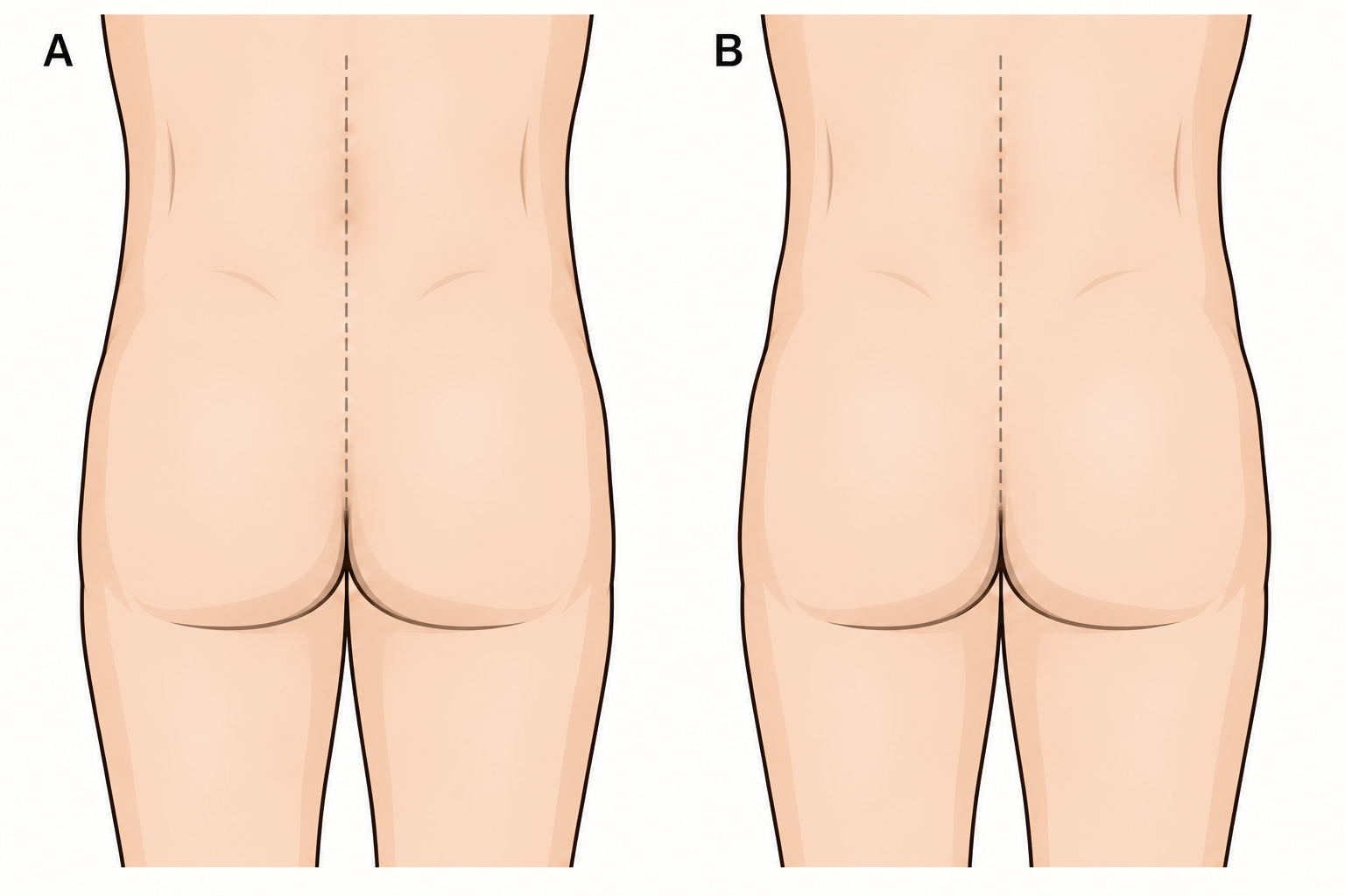
- 1 Classic Limberg: lower end on the midline
- 2 Mentes modification: lower end off the midline
Description of the drawing
A shows the classic Limberg plan, and B shows the Mentes modification. In the Mentes modification, the lower end of the area to be removed is moved away from the midline. The pale vertical dashed line shows the body's midline, and the dark dashed lines show the plan for the area to be removed and the flap; this plan is not an exact depiction of the scar after surgery.[16]
Results may vary from person to person with any surgical or interventional procedure. You are advised to discuss the procedure in detail with your doctor beforehand.
Illustrative drawing, not to scale. Source: Adhikari Sudhin et al. / Birat Journal of Health Sciences (2021), CC BY 4.0. Panels A and B of Figure 1 in the source article were adapted to a simple, high-resolution drawing of a person; the body was drawn as a male silhouette. The surgical planning lines were drawn on the web using coordinates. Panels C, D and E were removed; Turkish HTML explanations and leader lines were added. This adaptation is also licensed under CC BY 4.0.
Compared with leaving the wound open, the wound probably heals on average about six weeks sooner after flap surgery[1]; a flap may also reduce the chance of the disease returning, but the evidence for this is weak[1]. However, a stitched wound can open: in studies reported by the Dutch guideline, the wound opened in 17% of patients after flap surgery[1]; in studies reported by the German guideline, wound inflammation was reported in 8% to 50% of patients after the Karydakis method[2]. It is not known which method is best[1, 3, 8]; the Dutch guideline recommends using the flap with which the surgeon has the most experience, and the European Society of Coloproctology recommends choosing the method according to the surgeon's experience and the patient's preference[1, 8].
These operations are usually performed under a general or regional anaesthetic (from the waist down)[2]. According to the German guideline, after the Karydakis method the hospital stay is usually 0–3 days and time off work is 2–3 weeks[2]. Whether a drain (a thin tube that carries away fluid that collects) is placed in the wound is determined by the surgeon's preference and the individual patient[3, 8].
If the disease returns
The return of the disease (recurrence) is its reappearance after the wound has completely closed and symptoms have gone following treatment[1, 3]. Recurrences can also happen late: many are seen only after 1 year, or even more than 5 years[1]; in an analysis reported by the German guideline, nearly 30% of recurrences appeared more than four years after the first treatment[2]. Being of adolescent age, the number of openings and the size of the cavity are among the factors said to increase the likelihood of recurrence[3]; the European Society of Coloproctology adds obesity, smoking and previous surgery to these[8]. In one study, the disease returned more often in patients who smoked and whose openings were removed through small incisions[1].
Treatment of recurrent disease follows the same principles as treatment of disease appearing for the first time: if there is an abscess, it is drained; flap surgery may be needed for long-standing recurrent disease; the method is again chosen for the individual[3]. The Dutch guideline considers that people with complex or recurrent disease will benefit most from a flap[1]. The European Society of Coloproctology states that camera-assisted or other minor procedures may also be offered to selected patients with recurrent disease[8].
After surgery
Antibiotics are not routinely recommended after surgery[8]. If there are signs of infection, your doctor will decide whether they are needed[8].
Special dressing materials have not been shown to be better for wounds left open[3, 8]. According to the German guideline, these wounds should be prevented from sticking together and closing at the surface too early; showering several times a day with tap water of drinking-water quality is usually sufficient[2]. The German guideline recommends against treating the wound with a vacuum dressing[2]; the European Society of Coloproctology states that vacuum therapy may be used in selected patients for wounds that open after surgery[8]. The Italian consensus statement (2021), however, recommends vacuum therapy for wounds left open after extensive tissue removal; but in the study it cites, vacuum therapy produced faster healing only in the first two weeks, did not shorten the total healing time and prolonged the hospital stay[3]. A wound that does not heal after surgery and develops excessive, raised wound tissue (hypergranulation) should preferably be treated jointly by a surgeon and a wound care nurse[1].
Microscopic examination of the removed tissue is not needed for every patient[8]; the German guideline reminds readers that cancer can develop, although very rarely, and states that tissue examination is mandatory when cancer is suspected[2].
Returning to work or school depends on the procedure performed, the speed of healing and the type of work[4]. In studies pooled by the Dutch guideline, return to work after flap surgery was on average 5–6 days earlier than after surgery in which the wound was left open[1]; the German guideline states that time off work is 2–3 weeks after the Karydakis method and an average of one month when the wound is left open[2]. These are study averages; discuss the time that applies to you with your surgeon.
There is no single return-to-work time for the Limberg flap either. In a prospective case-control study from 2022 conducted in Turkey, the Limberg group's return to normal life and work was found to take an average of about 12 days[12]. In a randomised study from 2015 involving 91 patients, the modified Limberg group's return to work or school was an average of about 21 days[11]. The studies involved different patient groups and assessed different outcomes; these times are not a return date or a guarantee of an outcome for you.
If returning to work or school early is important to you, mention this at your examination. For limited disease, minor procedures such as Gips/pit picking or cleaning the channel using a camera may be considered; healing and time off work may be shorter[1, 2, 3]. However, they are not suitable for every form of the disease; the possibility of another procedure or the disease returning should also be discussed together when choosing a method[1, 2, 8].
The long-term rate at which the disease returns varies by method. In an analysis including more than 89,000 patients, the 10-year recurrence rate was found to be about 20% after surgery in which the wound was left open, about 3% after the Karydakis or Bascom “cleft lift” operation, about 11% after a Limberg or Dufourmentel flap, and up to 32% after surgery in which the wound was stitched on the midline[1, 2, 8]. NHS patient information describes surgery in which the wound is left open as the method with the lowest risk of recurrence[4]; however, the large analysis above found lower rates after the Karydakis or Bascom method and flap operations[1, 2, 8]. The rate of about 3% found for Karydakis and “cleft lift” in this large analysis is considerably lower than in individual studies with long-term follow-up after “cleft lift” (above, 11% to 24% at five years and beyond)[2]; the guidelines emphasise that there is no common classification of the disease, that the definition of recurrence varies between studies and that follow-up periods differ and are often short, so these rates cannot be directly compared[2, 8]. Recurrence has been reported after every method[1, 2, 3].
Hair removal: shaving or laser?
The Dutch guideline considers recommending against shaving between the buttocks with a razor[1]; razor shaving has been associated with more frequent recurrence of the disease in the long term[1, 2, 3]. The NHS also says not to shave the area unless your doctor recommends it[4].
The guidelines differ on whether laser hair removal (laser epilation) prevents the disease from returning. The Dutch guideline states that the evidence is very weak and that two studies gave opposite results; it recommends caution in recommending laser epilation after the first operation, but suggests considering it after discussing the advantages and disadvantages for people whose disease has returned several times and who have hair between the buttocks[1]. The Italian consensus statement recommends hair removal after surgery for patients with body hair, but states that the evidence for this is still weak[3]. The European Society of Coloproctology states that hair removal by any method after surgery is not essential to reduce recurrence: in a review it cites, recurrence was found in 9% after laser treatment, 19% of those who had no hair removal and 23% of those who removed hair by shaving or using a depilatory; but the society says further studies are needed before laser treatment can be recommended[8]. The German guideline, in its 2026 version, calls for laser epilation alongside surgery to be recommended to young adults aged 18–21; it states that the available evidence does not allow a recommendation for older people[2].
This recommendation in the German guideline is based on a study published after the Dutch guideline and the Italian consensus statement. In this study of 302 young people aged 11–21 who had previously been treated for an episode of pilonidal sinus, the disease returned significantly less often within one year in those who had laser epilation added to standard care (better hygiene and hair removal by shaving or depilatory cream): about 1 in 10 compared with 1 in 3[2, 9]. However, more than a third of the young people in the laser group did not attend the one-year follow-up[9]. The Dutch guideline noted that the comparison in the study design was with shaving; because shaving itself can increase recurrence, the results could misleadingly favour laser treatment[1].
Laser treatment usually cannot remove all the hair: at best, about 80% is removed; the procedure is painful, the degree of pain varies from person to person, and an average of six sessions is needed[1].
To protect yourself
The Dutch guideline considers giving the following advice to people with a pilonidal sinus that causes no symptoms, those who have symptoms but do not want treatment, and those in whose families pilonidal sinus is common[1]:
- after having your hair cut and removing body hair, clear away loose hairs that have fallen between the buttocks;
- do not shave between the buttocks with a razor;
- keep the area between the buttocks clean: the aim is to prevent loose hairs and debris from collecting there;
- lose weight if you have obesity;
- stop smoking;
- avoid sitting for long periods.
For people who are going to have or have had pilonidal sinus surgery other than abscess drainage, the Dutch guideline considers recommending stopping smoking before surgery and losing weight if they have obesity; after surgery, it considers recommending clearing away loose hairs, not shaving with a razor, keeping the area between the buttocks clean, not smoking and avoiding sitting for long periods[1]. The European Society of Coloproctology also recommends showering after a haircut and paying attention to hygiene[8].
These recommendations are based mostly on observational studies and expert opinion; although the evidence is not strong, they are recommended because they are harmless[1].
Sources
16
- Nederlandse Vereniging voor Heelkunde. Richtlijn Sinus pilonidalis. Richtlijnendatabase (Federatie Medisch Specialisten); modules 19.09.2022. Text: richtlijnendatabase.nl.
- Ommer A, Iesalnieks I, Doll D, Petersen S, Kahlke V, Schneider J, et al. S3-Leitlinie Sinus pilonidalis, Langfassung, Version 3.1. Deutsche Gesellschaft für Koloproktologie; AWMF-Registernummer 081-009. 2026. Text: register.awmf.org.
- Milone M, Basso L, Manigrasso M, Pietroletti R, Bondurri A, La Torre M, et al. Consensus statement of the Italian society of colorectal surgery (SICCR): management and treatment of pilonidal disease. Tech Coloproctol. 2021;25(12):1269–1280. doi: 10.1007/s10151-021-02487-8. PubMed: 34176001.
- NHS. Pilonidal sinus. Last reviewed: 16 November 2023. Text: www.nhs.uk.
- Sekhon Inderjit Singh HK, Meinero P, Campanile FC, Quddus A, Rajaganeshan R, Warusavitarne J, et al. The application of EPSiT in pilonidal sinus disease: an international Delphi consensus study endorsed by the Association of Laparoscopic Surgeons of Great Britain and Ireland (ALSGBI). Tech Coloproctol. 2025;29(1):155. doi: 10.1007/s10151-025-03191-7. PubMed: 40736584.
- Ouali M, Martin-Martin GP, Avdicausevic S, Alam A, Istok P, Brunenieks I, et al. International Society of Laser Proctology Position Paper on SiLaC and EPSiT/SiLaC in the Management of Pilonidal Sinus Disease. Lasers Surg Med. 2025;57(10):845–850. doi: 10.1002/lsm.70071. PubMed: 41078023.
- NHS. Sepsis. Last reviewed: 14 May 2026. Text: www.nhs.uk.
- Ojo D, Gallo G, Kleijnen J, Haas S, Danys D, Dardanov D, et al. European Society of Coloproctology guidelines for the management of pilonidal disease. Br J Surg. 2024;111(10):znae237. doi: 10.1093/bjs/znae237. PubMed: 39397672.
- Minneci PC, Gil LA, Cooper JN, Asti L, Nishimura L, Lutz CM, et al. Laser epilation as an adjunct to standard care in reducing pilonidal disease recurrence in adolescents and young adults: a randomized clinical trial. JAMA Surg. 2024;159(1):19–27. doi: 10.1001/jamasurg.2023.5526. PubMed: 37938854.
- Türk Dermatoloji Derneği. Hidradenitis süpürativa. Patient information leaflet. Text: turkdermatoloji.org.tr.
- Tokaç M, Dumlu EG, Aydın MS, Yalçın A, Kılıç M. Comparison of modified Limberg flap and Karydakis flap operations in pilonidal sinus surgery: prospective randomized study. Int Surg. 2015;100(5):870–877. doi: 10.9738/INTSURG-D-14-00213.1. PubMed: 26011208.
- Alkurt EG, Vardar YM, Tüzün İS. Comparison of Limberg flap and Karydakis flap repair in pilonidal sinus surgery: a prospective case-control study. Cureus. 2022;14(9):e28933. doi: 10.7759/cureus.28933.
- Pescatore R; Birnbaumer DM (reviewer). How to do procedural sedation and analgesia. MSD Manual Professional Edition. October 2024. Text: www.msdmanuals.com.
- Lim J, Shabbir J. Pilonidal Sinus Disease — A Literature Review. World J Surg Surgical Res. 2019;2:1117. Figure 1; full text accessed 3 October 2026. Text: www.surgeryresearchjournal.com.
- Hemmingsson O et al. Excision and suture in the midline versus Karydakis flap surgery for pilonidal sinus: randomized clinical trial. BJS Open. 2022;6(2):zrac007. Figure 2; full text accessed 3 October 2026. Text: pmc.ncbi.nlm.nih.gov.
- Adhikari Sudhin BN et al. Modified Limberg flap cover for recurrent sacrococcygeal pilonidal sinus: our experience at a medical college in Nepal. Birat Journal of Health Sciences. 2021;6(1):1274–1279. Figure 1; full text accessed 3 October 2026. Text: www.nepjol.info.
Medical review of the Turkish original: Op. Dr. Ömer Seyfullah Hasıripi, . This page is a translation checked by AI reviewers; the Turkish version prevails.
This page is for general information; it does not replace an examination. See a doctor about your symptoms.