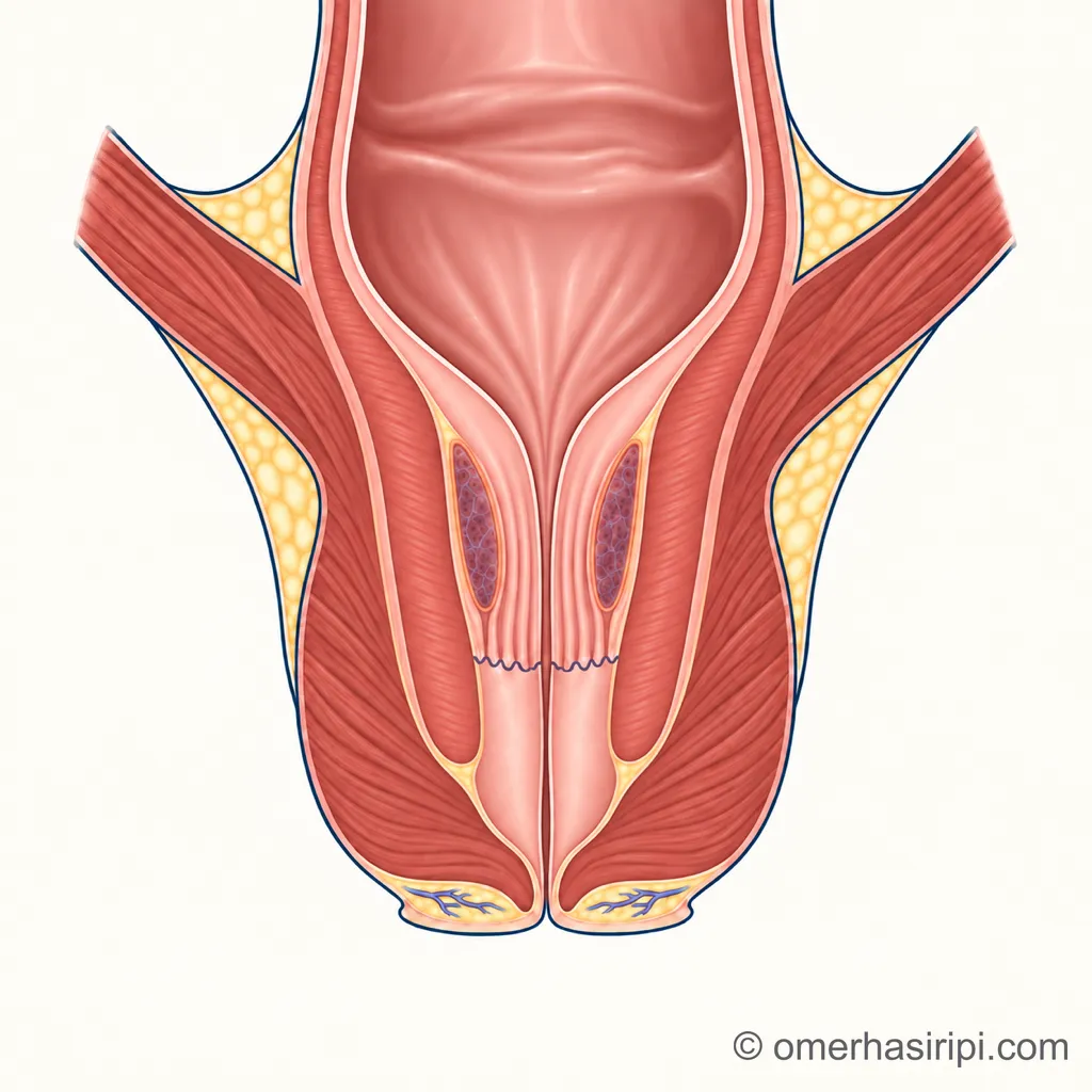
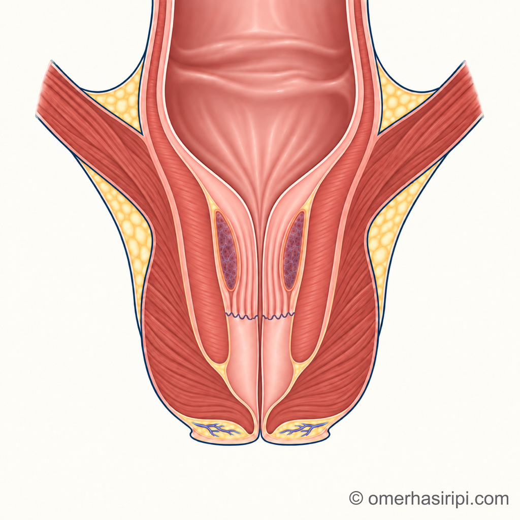
What are piles?
Spongy cushions rich in blood vessels are found on the inner surface of the anal canal. Everyone has these cushions, and they help the anus close tightly[3]. They fill with blood when you strain, helping to prevent stool, liquid and gas from leaking; about 15–20 per cent of the pressure that closes the anus comes from these cushions[4].
When the cushions become enlarged and cause symptoms, this is called haemorrhoidal disease, commonly known as piles[3]. Haemorrhoids that do not cause symptoms are not considered a disease, and it is recommended that they are not treated with procedures[3].
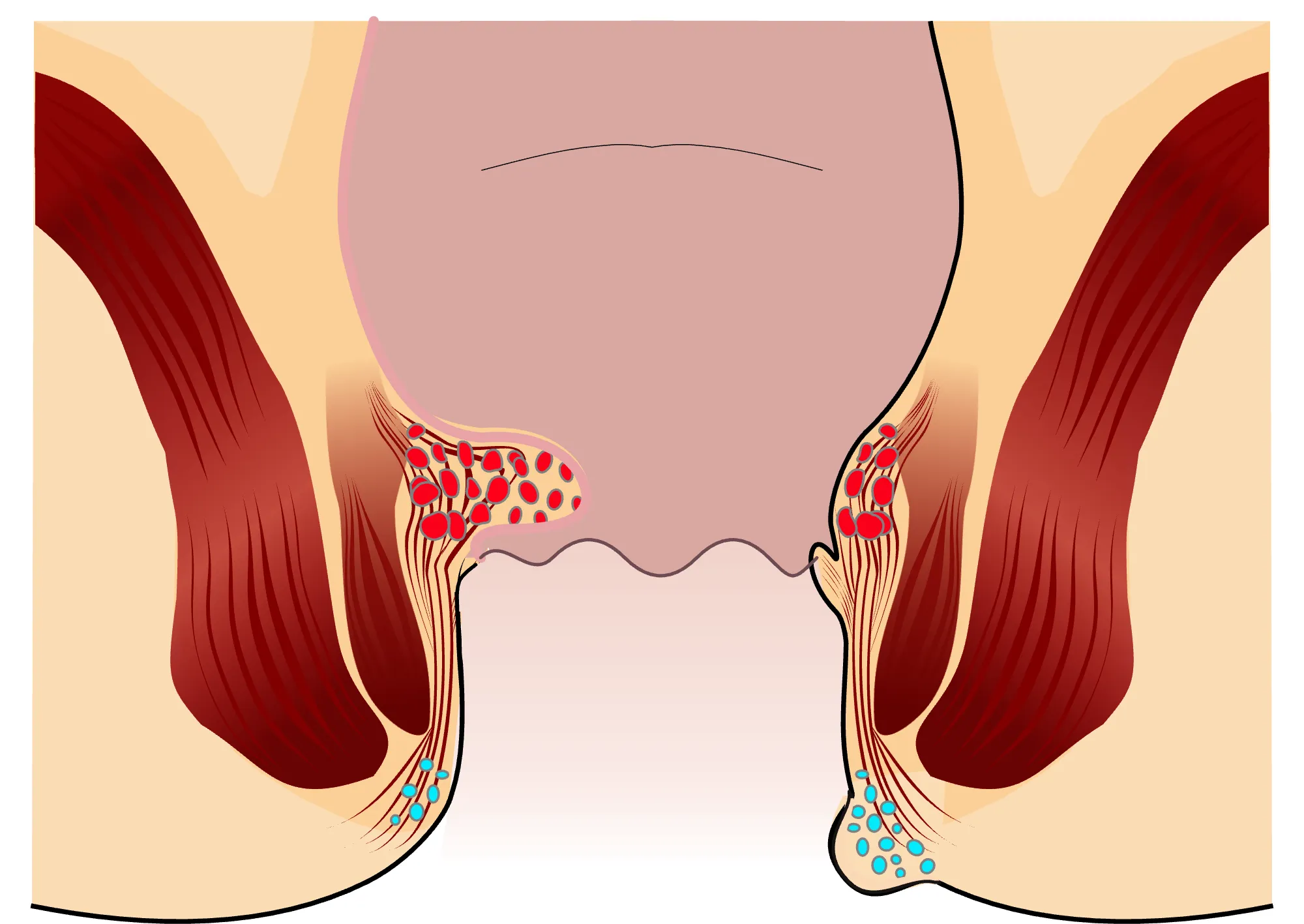
Enlarge
Swipe the drawing right or left to see the details. You can use the arrow keys on your keyboard.
- 1 Internal haemorrhoid
- 2 External haemorrhoid
- 3 Dentate line
Description of the drawing
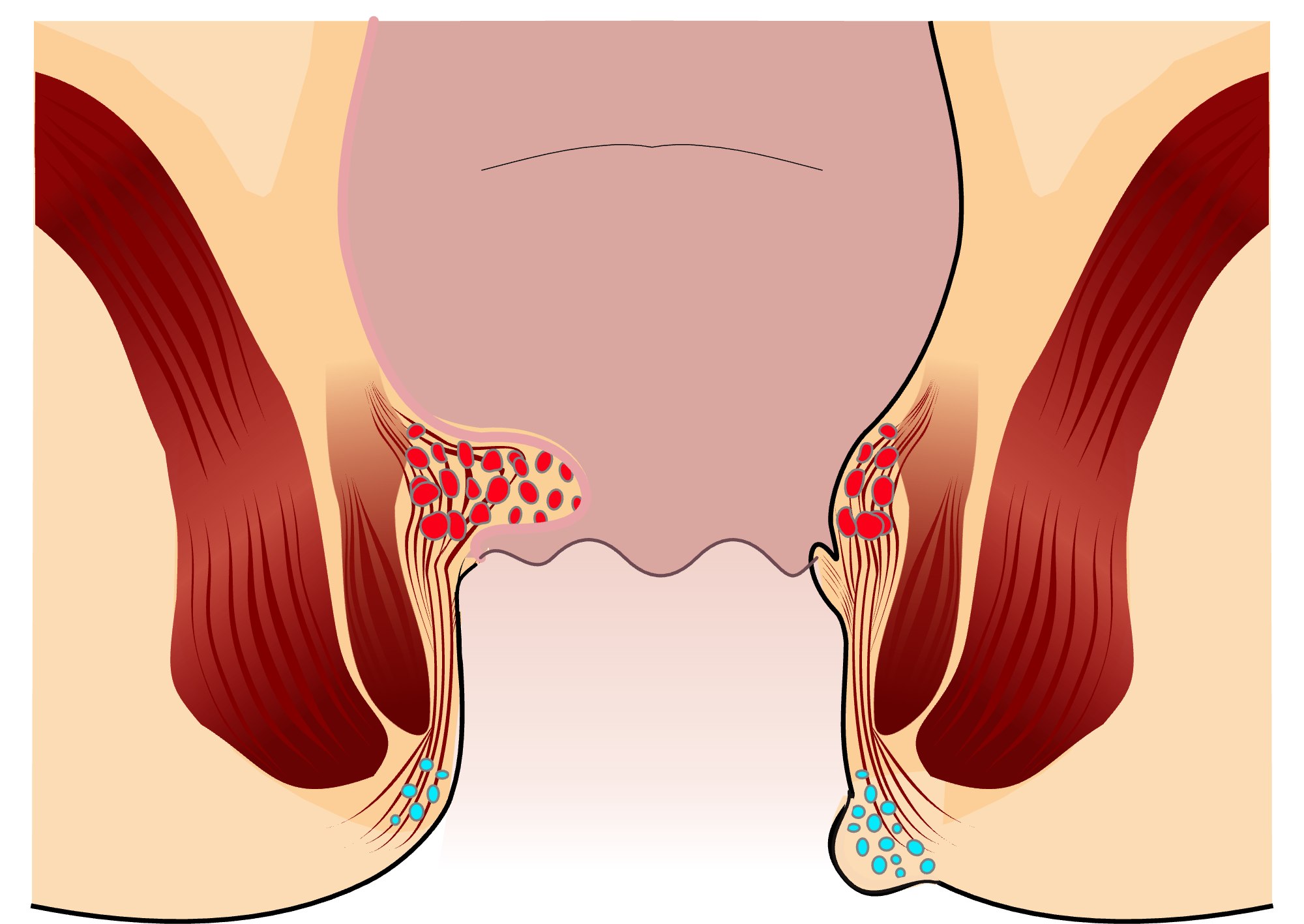
The dentate line separates internal from external haemorrhoids: internal haemorrhoids develop from cushions above this line and are covered by the inner lining of the bowel (mucosa); external haemorrhoids lie under the skin below the line, at the edge of the anal opening. The anal canal is drawn open to show the structures; it is normally closed.[5, 19]
Illustrative drawing, not to scale. Source: Armin Kübelbeck, CC BY 3.0. Source drawings: Piles Grade 1, Piles Grade 2, Analthrombose 01. The source drawing was adapted. Parts from the drawings “Piles Grade 1”, “Piles Grade 2” and “Analthrombose 01” (Wikimedia Commons) were combined, the internal haemorrhoid cushion was reshaped and an inner lining added, the colour inside the bowel was changed, and the inner surface below the dentate line was coloured a lighter shade; numbered labels were added.


What are the symptoms?
The most common symptom is painless, bright red bleeding during or immediately after a bowel movement; the blood is usually noticed on the stool or on toilet paper[2]. Swelling at the anus, prolapse, leakage that soils underwear, itching, skin irritation and discomfort may also occur[2].
Frequently recurring bleeding may, over time, lead to anaemia caused by iron deficiency; rarely, the bleeding may be severe enough to require hospital treatment and a blood transfusion[2].
Internal haemorrhoids usually present with painless bleeding, especially if they do not prolapse. With external haemorrhoids, pain can be marked, especially when a clot forms inside; prolapsed internal haemorrhoids can also cause pain and discomfort[5, 10]. If pain is severe or continuous, other causes such as an anal fissure and an abscess are also investigated[2]. Symptoms of piles can also occur in a similar way with other anal conditions, and the severity of symptoms does not always correspond to the size of the haemorrhoids[3].
Stages of piles
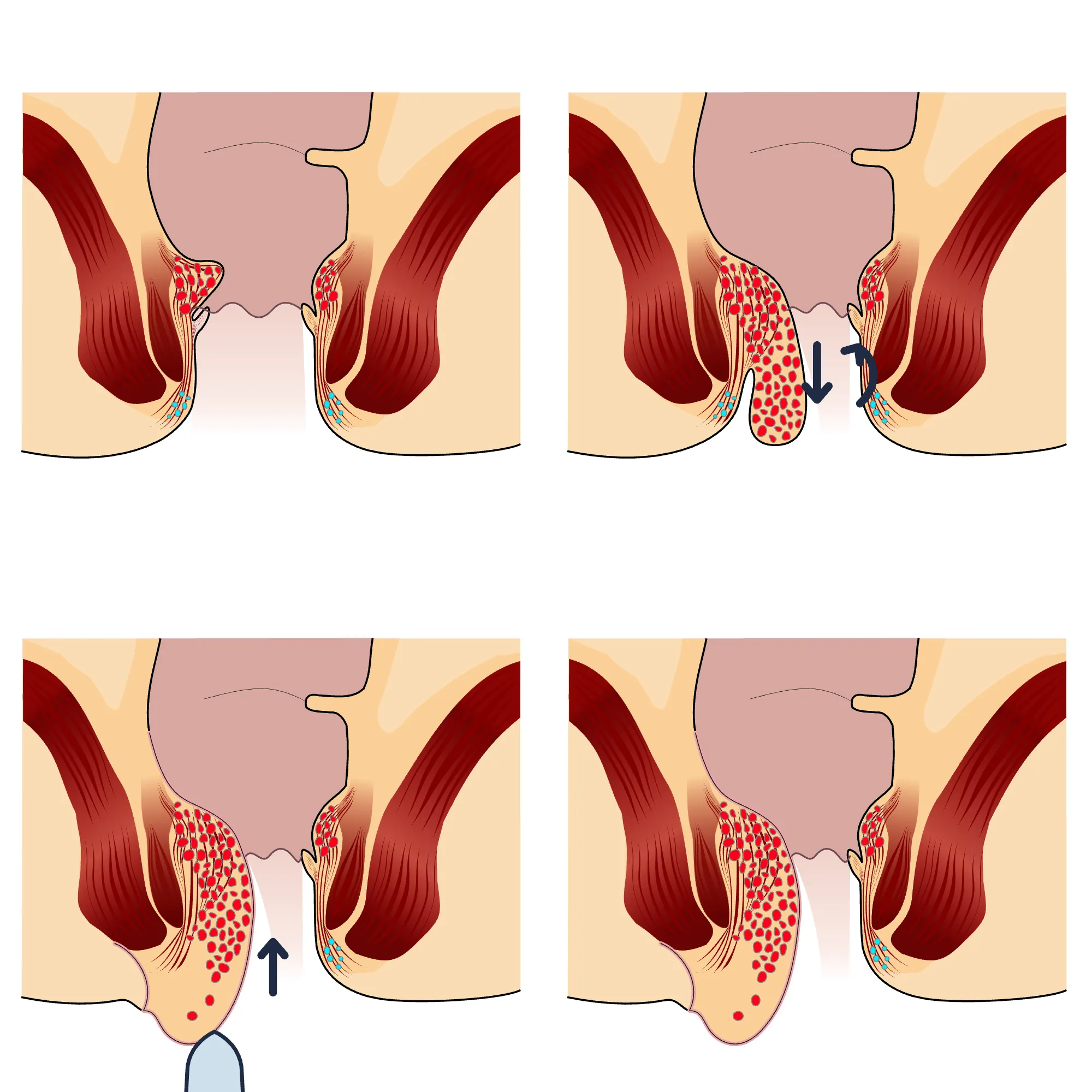
Doctors divide internal haemorrhoids into four stages according to whether and how they prolapse (the Goligher classification). The stage helps guide the choice of treatment[1].
Enlarge
Swipe the drawing right or left to see the details. You can use the arrow keys on your keyboard.
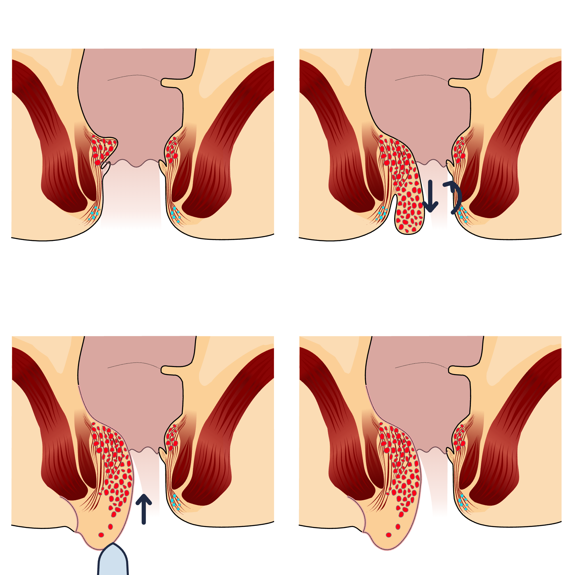
- 1 Stage 1: does not prolapse
- 2 Stage 2: prolapses when straining and goes back in on its own
- 3 Stage 3: prolapses and can be pushed back in by hand
- 4 Stage 4: remains outside all the time and cannot be pushed back in by hand
Description of the drawing
The four panels compare the stages of internal haemorrhoids; a description of each stage appears beside the numbers. The anal canal is drawn open to show the structures; it is normally closed.[4]
Illustrative drawing, not to scale. Source: Armin Kübelbeck, CC BY 3.0. Source drawings: Piles Grade 1, Piles Grade 3, Piles Grade 4. The source drawing was adapted. The drawings “Piles Grade 1”, “Piles Grade 3” and “Piles Grade 4” (Wikimedia Commons) were combined in four panels, the colour inside the bowel was changed, the inner surface below the dentate line was coloured a lighter shade, and arrows and a gloved finger were added; numbered labels were added.


When should bleeding from the anus be investigated?
Bleeding is the most common symptom of piles, but the colour or amount of blood does not show for certain where the bleeding comes from. For this reason, bleeding from the anus should be investigated by examination[3].
The diagnosis is made by assessing the symptoms and examining the anus[2]. When needed, piles and other anal conditions are assessed by looking inside the anus with an endoscopic procedure such as a colonoscopy or with an anoscope[1, 5, 13]. If the examination cannot be performed because of pain, the doctor may plan an assessment under anaesthesia[15]. A colonoscopy also investigates other causes of bleeding in the large bowel[5, 13]. According to the German guideline, however, the stage of piles cannot be reliably determined during colonoscopy[3]; findings in the anal canal are also identified more accurately with anoscopy than with colonoscopy[3, 13].
Who needs an examination of the large bowel with a camera (colonoscopy) depends on age, history and examination findings[3, 5]. The Italian Society of Colorectal Surgery recommends colonoscopy for every patient with piles and bleeding from the anus to rule out other bowel diseases[2]; the American guideline recommends it for selected patients[5].
Enlarge
Swipe the drawing right or left to see the details. You can use the arrow keys on your keyboard.
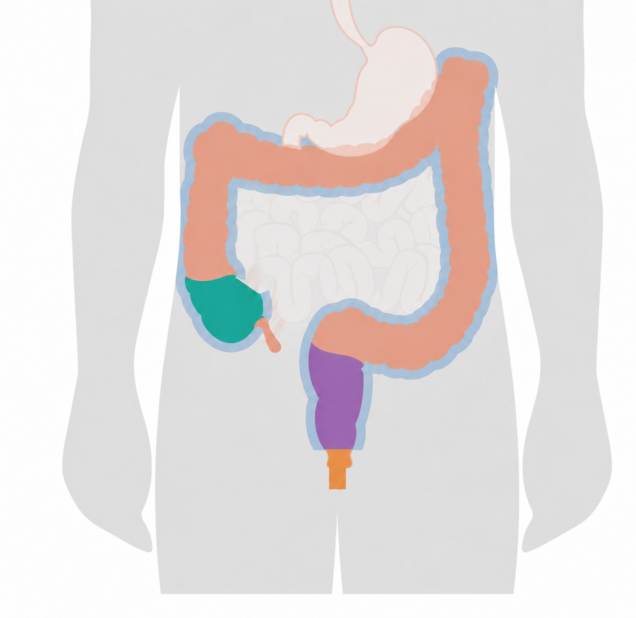
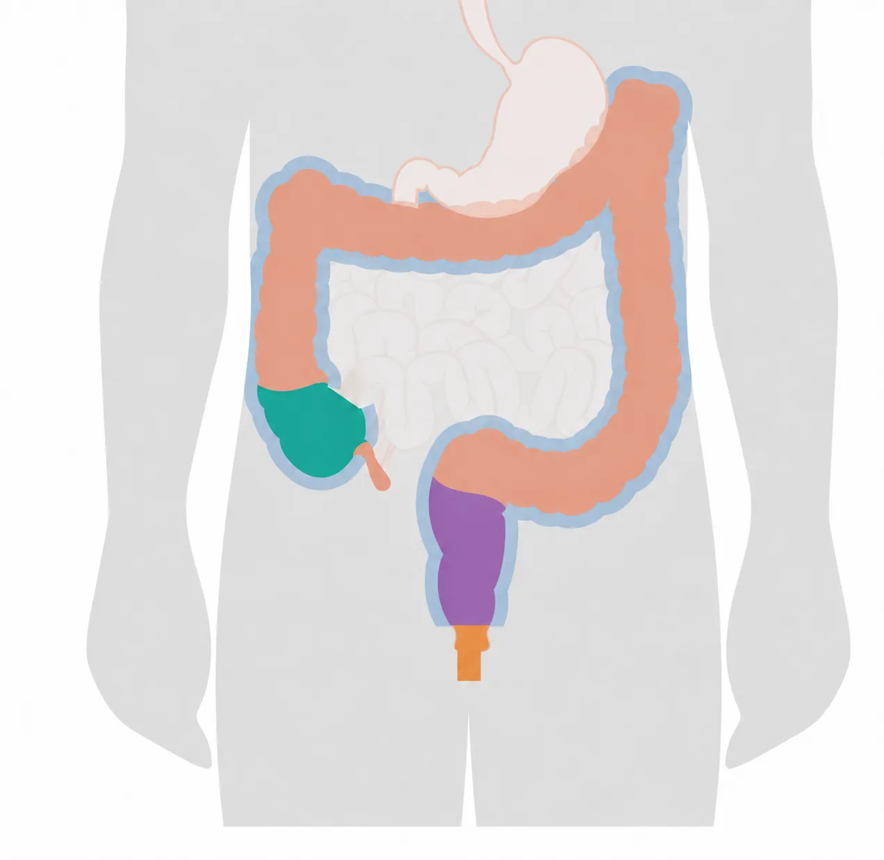
- 1 Orange: the anal canal examined by anoscopy
- 2 Blue band: the area examined by colonoscopy
- 3 Blind end of the large bowel (caecum, green)
- 4 Rectum (purple)
Description of the drawing
The blue band shows the area examined by colonoscopy: the large bowel from the rectum to the blind end of the large bowel (caecum). The orange section is the anal canal, which anoscopy examines; the colonoscope also enters through it. The green section is the caecum, and the purple section is the rectum. The coloured boundaries are illustrative.[5, 18]
Illustrative drawing, not to scale. Source: Cancer Research UK / Wikimedia Commons, CC BY-SA 4.0. The instrument, magnification window, tumour and English text were removed, the small bowel was faded, two coloured areas were added and the image was cropped; numbered labels were added. The caecum and rectum were coloured separately; the gap in the blue band where the rectum and sigmoid colon meet was closed, and the band was ended where the rectum ends. This adaptation is also licensed under CC BY-SA 4.0.

In the following situations, it is particularly important to make sure that the bleeding is not caused by something else, such as bowel cancer. The Italian statement considers colonoscopy essential in most of these situations[2]; the extended version of the European guideline also calls for particular care to rule out bowel cancer in some of them[4]:
- older age[2]: according to the German guideline, most studies recommend endoscopic examination of the bowel from age 40 in people with bleeding from the anus[3]; the extended version of the European guideline draws particular attention to those over 50[4],
- a personal or family history of bowel cancer (the European guideline mentions first-degree relatives, meaning parents, siblings and children), or a precancerous polyp with advanced features previously found in you (advanced adenoma)[2, 4],
- a history of inflammatory bowel disease[2],
- a change in bowel habits[2, 4],
- recent significant weight loss[2, 4],
- blood mixed with the stool[4],
- a test showing anaemia caused by iron deficiency or a positive test for hidden blood in the stool[2],
- another unusual finding around the anus on examination[4].
People with piles do not have a higher risk of bowel cancer than the general population[3]. But only an examination and, when needed, a colonoscopy can show whether the bleeding is caused by piles or something else[3]. See a doctor if your symptoms worsen or do not improve despite measures you take at home, if piles recur frequently, or if you notice a change around the anus that seems unusual to you[6].
Which department treats piles?
The general surgery department treats piles. Your family doctor or workplace doctor can carry out the initial assessment and refer you to the appropriate specialist if needed. The gastroenterology department also assesses piles and provides some treatments[14].
What can you do at home?
At every stage of piles, the first step in treatment is lifestyle measures and basic treatment[1]. These measures aim to relieve symptoms; they do not restore enlarged cushions to their original state[2].
- Fibre: Getting fibre every day from food or a supplement reduces the risk of bleeding and of symptoms continuing; its benefit for prolapse, pain and itching is more limited[5]. Daily fibre needs vary with energy intake: about 14 grams of fibre are recommended for every 1,000 calories. For example, this is about 28 grams for a 2,000-calorie diet. Vegetables, fruit, pulses and whole grains are sources of fibre; if needed, your doctor or dietitian may recommend a fibre supplement[11].
- Water and movement: Enough fluid helps fibre soften the stool. AGA patient information recommends 6–8 glasses of fluid a day; however, the daily amount varies according to your health, activity and the weather. If you have a fluid restriction because of heart or kidney disease, follow your doctor’s advice[11, 10]. Include activities such as regular walking in your daily life. For general adult health, 150–300 minutes of moderate-intensity activity a week is recommended; this is not a treatment dose specifically for haemorrhoids[12].
- Toilet habits: Do not put off the urge to open your bowels. Avoid straining and sitting on the toilet for a long time; get up when you have finished[5, 10]. AGA patient information recommends not sitting on the toilet for longer than five minutes[10].
- Creams and suppositories: These may temporarily relieve symptoms; a benefit from long-term use has not been shown, and they may cause a skin reaction[2]. They do not treat piles; they only relieve symptoms[3].
- Medicines known as blood vessel strengtheners: These may help reduce symptoms; however, their actual effects are uncertain because the quality of the research is limited[1, 2]. Consult your doctor before taking medicine.
- Warm sitz bath: This is a traditional and frequently recommended method; however, comparative studies in which patients are randomly assigned to groups are lacking to establish its role in pain from piles[2].
Treatment with minor procedures
If basic treatment does not reduce symptoms sufficiently, less invasive procedures are considered before surgery: rubber band ligation, sclerotherapy and infrared light treatment[1].
Rubber banding (band ligation)
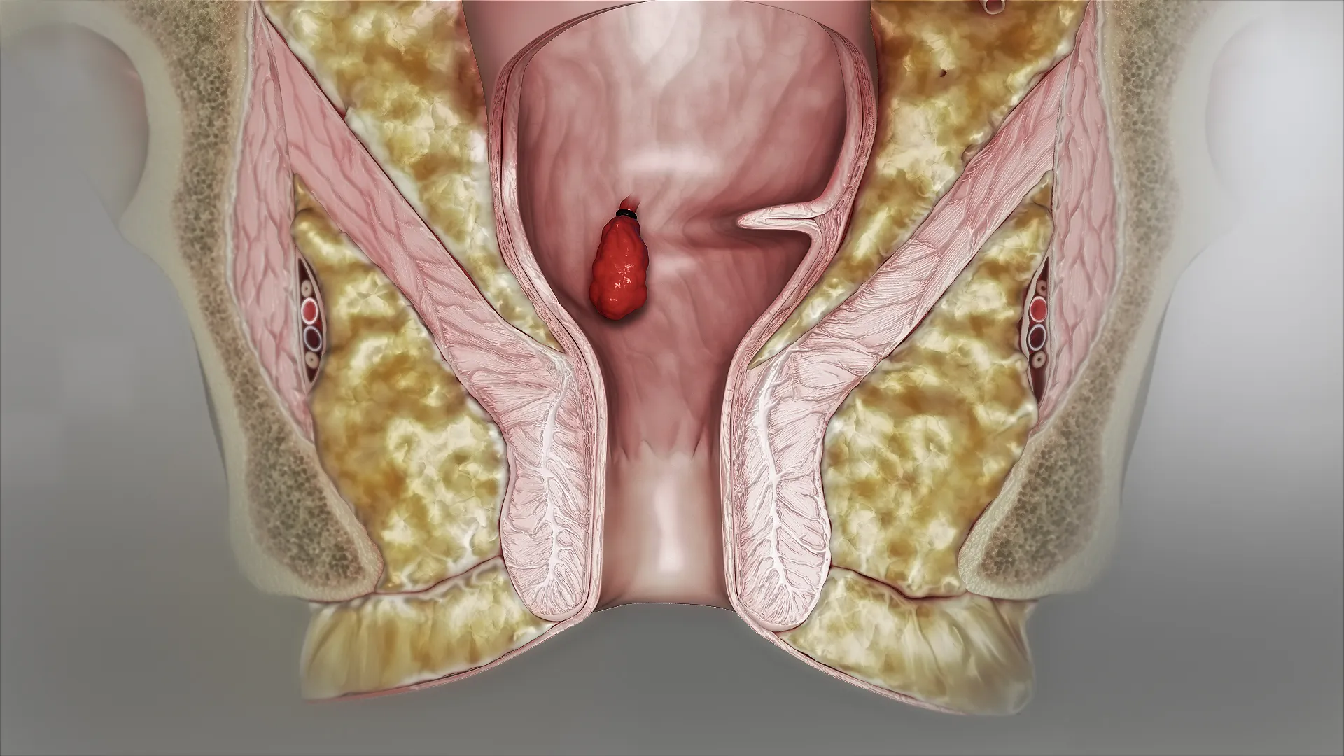
The base of the haemorrhoid cushion is tied with a small rubber band; the banded tissue dries up and falls off over time[2]. It is used for stages 1 and 2, and in selected patients with stage 3, and can be repeated if needed[5]. It is considered the most effective option among the minor procedures[5].
Enlarge
Swipe the drawing right or left to see the details. You can use the arrow keys on your keyboard.
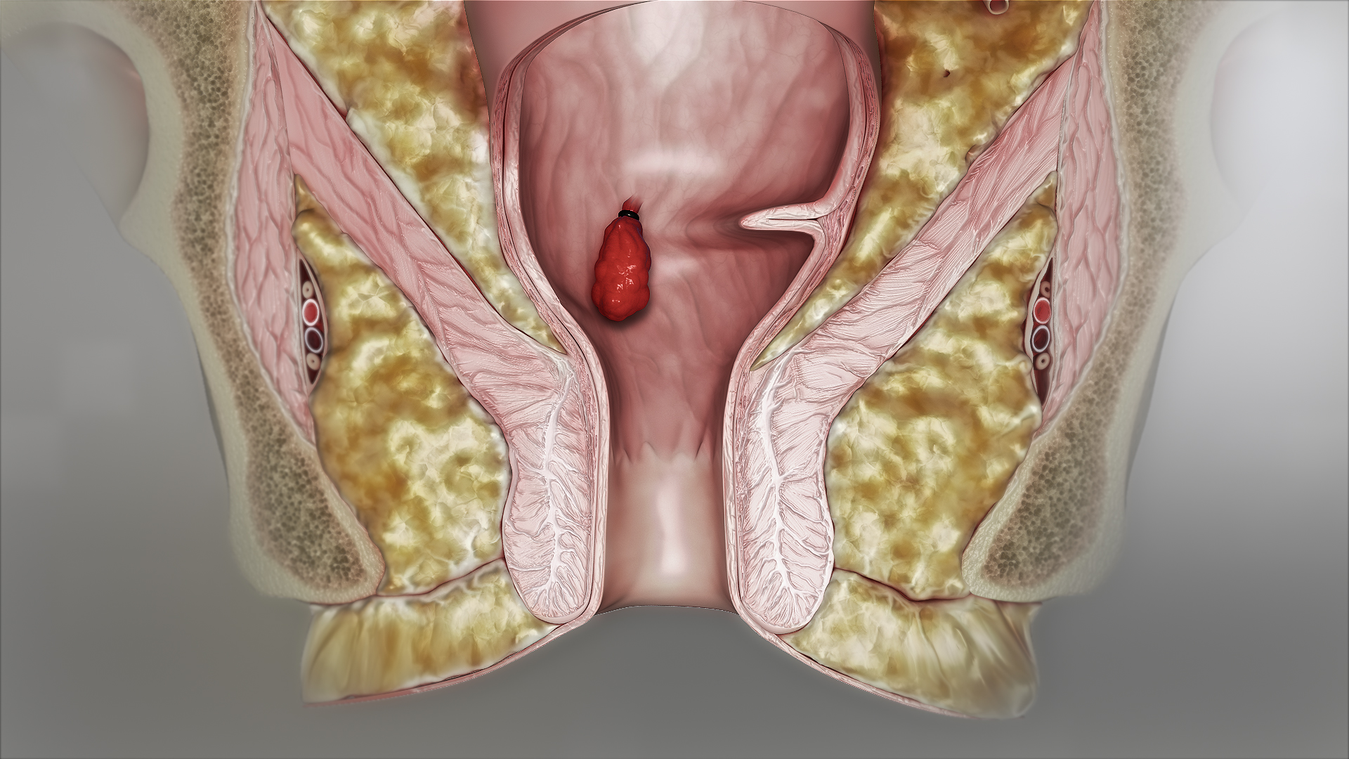
- 1 Rubber band tightening around the base of the haemorrhoid
- 2 Internal haemorrhoid tissue tied with a rubber band
Description of the drawing
The rubber band tightens around the base of the internal haemorrhoid above the dentate line; the part whose blood supply is cut off falls off over time. In the drawing, the tissue tied with the band appears connected to the bowel wall by a narrow neck; the dentate line is not shown. The drawing shows only the stage at which the band is placed; the anal canal is shown open so that its inside can be seen.[5]
Results may vary from person to person with any surgical or interventional procedure. You are advised to discuss the procedure in detail with your doctor beforehand.
Illustrative drawing, not to scale. Source: Scientific Animations (2018), CC BY-SA 4.0. The original source image was retained; Turkish numbered descriptions were added.

The most common side effects are pain and bleeding[3]. A small amount of bleeding may occur after the procedure[7]. More noticeable bleeding can occur, usually 10 to 14 days after the procedure, as the banded tissue falls off[5, 7]; this risk is higher in people taking a blood thinner or an anti-clotting medicine[2, 3]. If you have unbearable pain immediately after banding, tell your doctor immediately: the band may have been placed too low and may need to be removed[3, 4]. Although very rare, serious complications have been reported, including heavy bleeding, a severe infection (sepsis) that can develop around the anus and in the pelvis (the area in the lower part of the abdomen containing the anus and urinary tract) and rapidly become worse, and, in very exceptional cases, death[2, 3].
Sclerotherapy
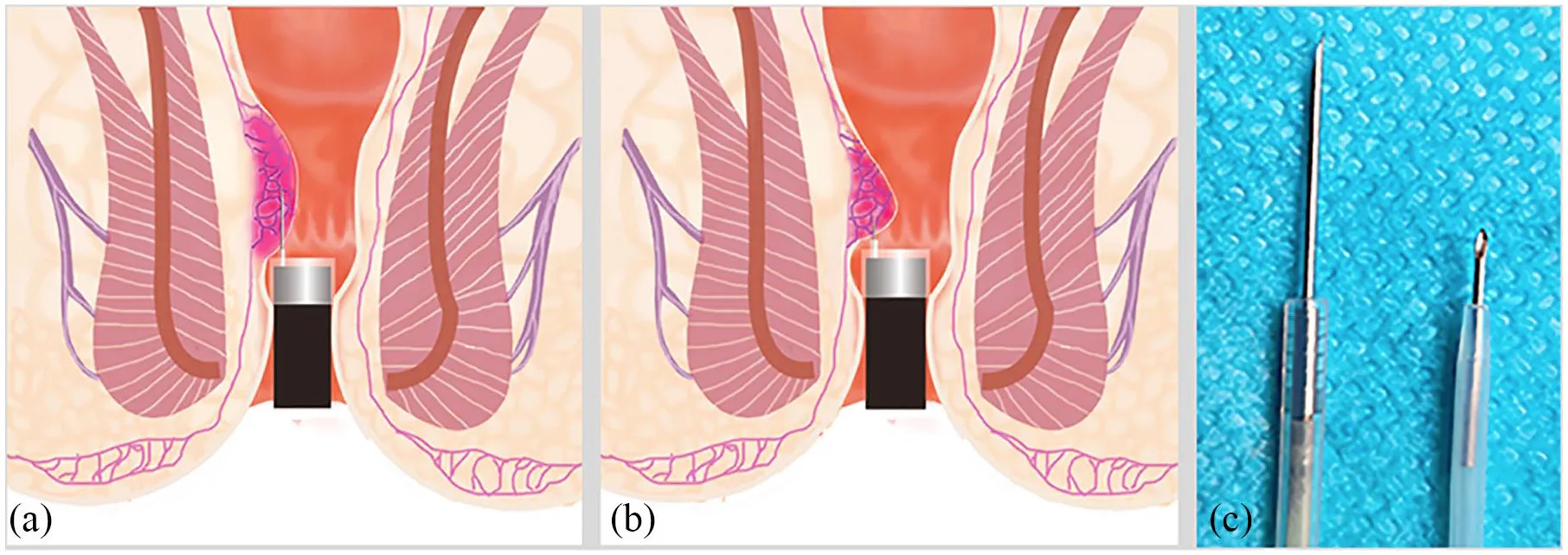
A medicine that hardens blood vessels is injected into the haemorrhoidal tissue. The European and German guidelines state that it can be used for grades 1–2[1, 3]; the Italian statement also includes grade 3[2]. According to the German guideline, the injection is given in an area without pain sensation[3]; nevertheless, in one study most patients reported feeling pain during the injection[2].
Enlarge
Swipe the drawing right or left to see the details. You can use the arrow keys on your keyboard.
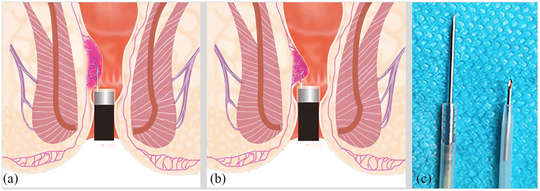
- 1 Internal haemorrhoid cushion
- 2 Injection needle
- 3 Cap at the tip of the endoscope
Description of the drawing
An example of cap-assisted endoscopic sclerotherapy: the cap at the tip of the endoscope makes it easier to see the area of the internal haemorrhoid. The hardening agent (sclerosant) is injected into the tissue beneath the mucosa; the aim is to form connective tissue and fix the tissue in place. The diagrams on the left and in the centre show the procedure with a long and a short needle, while the photograph on the right shows these needles. The source is a research protocol; this drawing cannot be used to conclude that one needle is better than the other.[5, 22]
Results may vary from person to person with any surgical or interventional procedure. You are advised to discuss the procedure in detail with your doctor beforehand.
Illustrative drawing, not to scale. Source: Xia Wu et al. (2020), Figure 3, CC BY 4.0. Source drawings: Original Figure 3. The original image was preserved; Turkish HTML pins and explanations were added.
Treatment with infrared light (infrared coagulation)
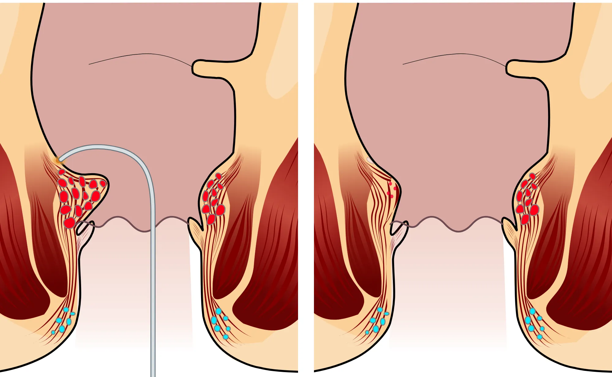
It can be used as an option particularly for bleeding grade 1 haemorrhoids[1]. The tissue is shrunk by heat produced by infrared light; this procedure is different from treatments using a laser[5, 16].
Enlarge
Swipe the drawing right or left to see the details. You can use the arrow keys on your keyboard.
- 1 Infrared application tip
- 2 Internal haemorrhoid
- 3 Dentate line (the natural boundary inside the anal canal)
- 4 Healing mark
- 5 Shrunken internal haemorrhoid
Description of the drawing
On the left, the application tip touches immediately above the internal haemorrhoid; the orange area represents the effect of heat. On the right is an illustrative view of the same area after some time: the tissue has shrunk, leaving a pale healing mark. The contact point and the mark are above the dentate line. Only the tip of the instrument is drawn; the other instruments used in the procedure are not shown. The single contact point is an example; the tip is also moved to neighbouring areas. The drawing is schematic and not to scale. The canal is drawn open for visibility; it is normally closed.[3, 5, 23]
Results may vary from person to person with any surgical or interventional procedure. You are advised to discuss the procedure in detail with your doctor beforehand.
Illustrative drawing, not to scale. Source: Armin Kübelbeck, CC BY 3.0. Source drawings: Piles Grade 1. The source drawing was adapted. The colours were adjusted, the cross-section was cropped and two panels were created. A curved infrared application tip and a small area representing the effect of heat were added. Shrunken internal haemorrhoid tissue and a pale healing mark on the surface were shown in the second panel. Adapted from Armin Kübelbeck’s “Piles Grade 1” drawing under the CC BY 3.0 licence.
Pain may occur during or after these procedures[2, 3]; the German guideline calls for the possibility of pain and bleeding with rubber band ligation to be discussed before the procedure[3]. If you take a blood thinner or a medicine that prevents clotting, be sure to tell your doctor: whether the medicine should be paused before the procedure and, if so, how this should be done is planned according to national recommendations[1]; some procedures can also be performed without pausing the medicine[2, 3]. Do not stop taking the medicine on your own. Inflammatory bowel disease, radiotherapy to the pelvis (the area containing the organs in the lower part of the abdomen), a disease or medicine that suppresses the immune system, a bleeding disorder, diabetes and pregnancy are also things you need to tell your doctor about before the procedure[1, 2].
When is surgery needed?
Surgery is considered particularly at an advanced grade or when other treatments do not give sufficient results. The grade alone does not determine the decision: surgery may also be considered for selected patients at an early grade if previous treatments have not worked or are unsuitable[5, 13]. The presence of both internal and external haemorrhoids, the severity of symptoms, the patient’s preference and general health are considered together[1, 5].
Main methods
- Removal of haemorrhoidal tissue (haemorrhoidectomy): The 2020 Italian consensus statement recommends this classic method as the first choice when surgery is needed for grade 3 and 4 haemorrhoids that cause symptoms[2]. The wound may be left open (Milligan–Morgan) or closed with stitches (Ferguson); therefore, not all haemorrhoidectomies are “open surgery”[5]. It causes more pain than other methods in the first few days[2, 3].
- Stapled procedure: Compared with classic surgery, there is less pain, the hospital stay is shorter and patients return to work sooner; however, recurrence of haemorrhoids and the need for another operation are more common[2]. The German guideline recommends against using this method for grade 4 haemorrhoids[3].
- Haemorrhoidal artery ligation (HAL or THD), with lifting of prolapsed tissue if needed: HAL stands for Hemorrhoidal Artery Ligation (tying off the haemorrhoidal arteries); THD stands for Transanal Hemorrhoidal Dearterialization (reducing blood flow in the arteries that supply the haemorrhoids by entering through the anus)[5, 17]. It is considered an option for grades 2 and 3[1, 2]. There may be less pain than with classic surgery; however, recurrence may be more common[2, 5]. The German guideline recommends against Doppler-guided haemorrhoidal artery ligation (DGHAL) for grades 3 and 4 because its recurrence rate is higher than with classic operations[3].
- Newer methods such as laser and radiofrequency: The European guideline states that these methods can be used, but that the level of evidence is low[1]; the German guideline makes no recommendation for laser methods[3]. Laser methods differ from one another, and different outcomes have been reported[2, 3]. For the method in which a laser is applied inside the haemorrhoidal cushion (laser haemorrhoidoplasty), comparative studies have reported less pain than with classic open surgery; in a single study of this method, haemorrhoids recurred in 39% of patients with grade 2 and 33% of those with grade 3 during an average follow-up of 5.4 years[2]. The laser haemorrhoidoplasty consensus published in 2024 identifies patients with symptomatic grade 2 and 3 haemorrhoids as the main patient group for using this method alone. The consensus recommendations do not mean that laser is superior to other methods for every patient; recurrence and the need for additional procedures should be discussed beforehand[16].
No method is best for every patient. There are few high-quality studies comparing the methods, and their results cannot be fully compared with one another[1, 3]. Haemorrhoids can recur after any method[2, 3].
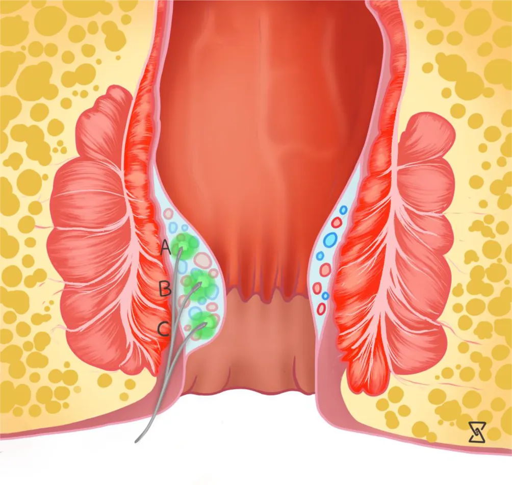
Diagram of laser haemorrhoidoplasty
Enlarge
Swipe the drawing right or left to see the details. You can use the arrow keys on your keyboard.
- 1 Entry route of the laser fibre
- 2 Laser fibre passing beneath the mucosa
- 3 Internal haemorrhoid cushion
- 4 Dentate line
Description of the drawing
Diagram of laser haemorrhoidoplasty (LHP): a thin fibre is advanced through a small entry at the edge of the anus into the area beneath the mucosa within the internal haemorrhoidal cushion. Laser energy is applied to this area; the aim is for the tissue to shrink and connective tissue to form over time. A, B and C in the source drawing are different treatment positions of the same fibre: A is above the dentate line, while B and C are in the upper and lower parts of the haemorrhoidal cushion; they are not three separate fibres used at the same time. The anal canal is drawn open to show the structures; it is normally closed.[13, 16, 21]
Results may vary from person to person with any surgical or interventional procedure. You are advised to discuss the procedure in detail with your doctor beforehand.
Illustrative drawing, not to scale. Source: Peter C. Ambe (2026), Figure 3, CC BY 4.0. Source drawings: Ambe 2026 Figure 3, the LHP recommendations from which this figure was taken (Ambe et al., 2025). The original image was preserved; Turkish HTML pins and explanations were added.
Diagram of THD and mucopexy
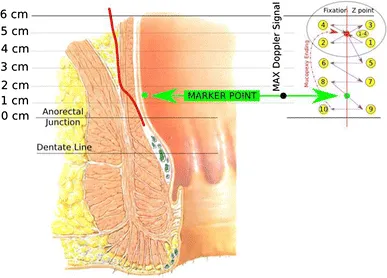
- 1 Internal haemorrhoid cushion
- 2 Ligation site identified by Doppler (marker point)
- 3 Diagram of the stitches that lift the mucosa
- 4 Dentate line (Dentate Line)
Description of the drawing
THD diagram: Doppler is used to locate the artery supplying the haemorrhoid, and the vessel is tied off with a stitch. If there is prolapse, a series of stitches that lifts the mucosa (mucopexy) may be added; the small diagram at the top right shows this series of stitches. In THD, the mucopexy stitch is stopped at the upper end of the internal haemorrhoidal cushion; the cushion is not included in this stitch. The small yellow numbers at the top right show the order in which the Z-shaped fixation stitch and the mucopexy stitch that follows it pass through the tissue in the source diagram; they are separate from the numbers 1–4 in the explanation. English terms in the drawing: Anorectal Junction, the point where the rectum joins the anal canal; MAX Doppler Signal, the strongest Doppler sound; Fixation Z point, the fixation point for the Z stitch; Mucopexy Ending, the end of the mucopexy. The distances in the source drawing belong to the diagram of this technique; they are not fixed surgical measurements for every patient.[5, 17]
Results may vary from person to person with any surgical or interventional procedure. You are advised to discuss the procedure in detail with your doctor beforehand.
Illustrative drawing, not to scale. Source: Carlo Ratto (2014), Figure 5, CC BY 2.0. Source drawings: Original Figure 5. The original image was preserved; Turkish HTML pins and explanations were added.
Diagram of closed haemorrhoidectomy
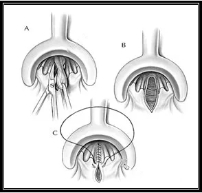
- 1 A: Removal of haemorrhoidal tissue
- 2 B: Wound after removal
- 3 C: Closure of the wound with stitches
Description of the drawing
Stages of closed haemorrhoidectomy: A shows removal of the haemorrhoidal tissue, B shows the wound after removal, and C shows closure of the wound with stitches. This drawing shows the Ferguson method. In open haemorrhoidectomy (Milligan–Morgan), the wound is not closed with stitches; it is left open. The open and closed methods are not two stages performed one after the other on the same patient.[5, 20]
Results may vary from person to person with any surgical or interventional procedure. You are advised to discuss the procedure in detail with your doctor beforehand.
Illustrative drawing, not to scale. Source: Andrea C. Bafford and Ronald Bleday (2008; via UCSF), CC BY 3.0. The original BMP file was converted to PNG without pixel loss; English text, two arrows and a spellcheck mark were removed; Turkish HTML pins and explanations were added.
A painful swelling that appears suddenly at the anus
A painful, usually bluish swelling that appears suddenly at the edge of the anus may be caused by a clot forming in a blood vessel beneath the skin[1, 2]. The European and German guidelines use the term “perianal thrombosis” (a clot at the edge of the anus) to distinguish this from haemorrhoids[1, 3]; the Italian and American guidelines call it “external haemorrhoid thrombosis”[2, 5]. Even without treatment, pain usually decreases within 2–3 days and the clot is gradually absorbed over a few weeks; painkillers and medicines that soften the stool may help[2].
Cross-sectional view
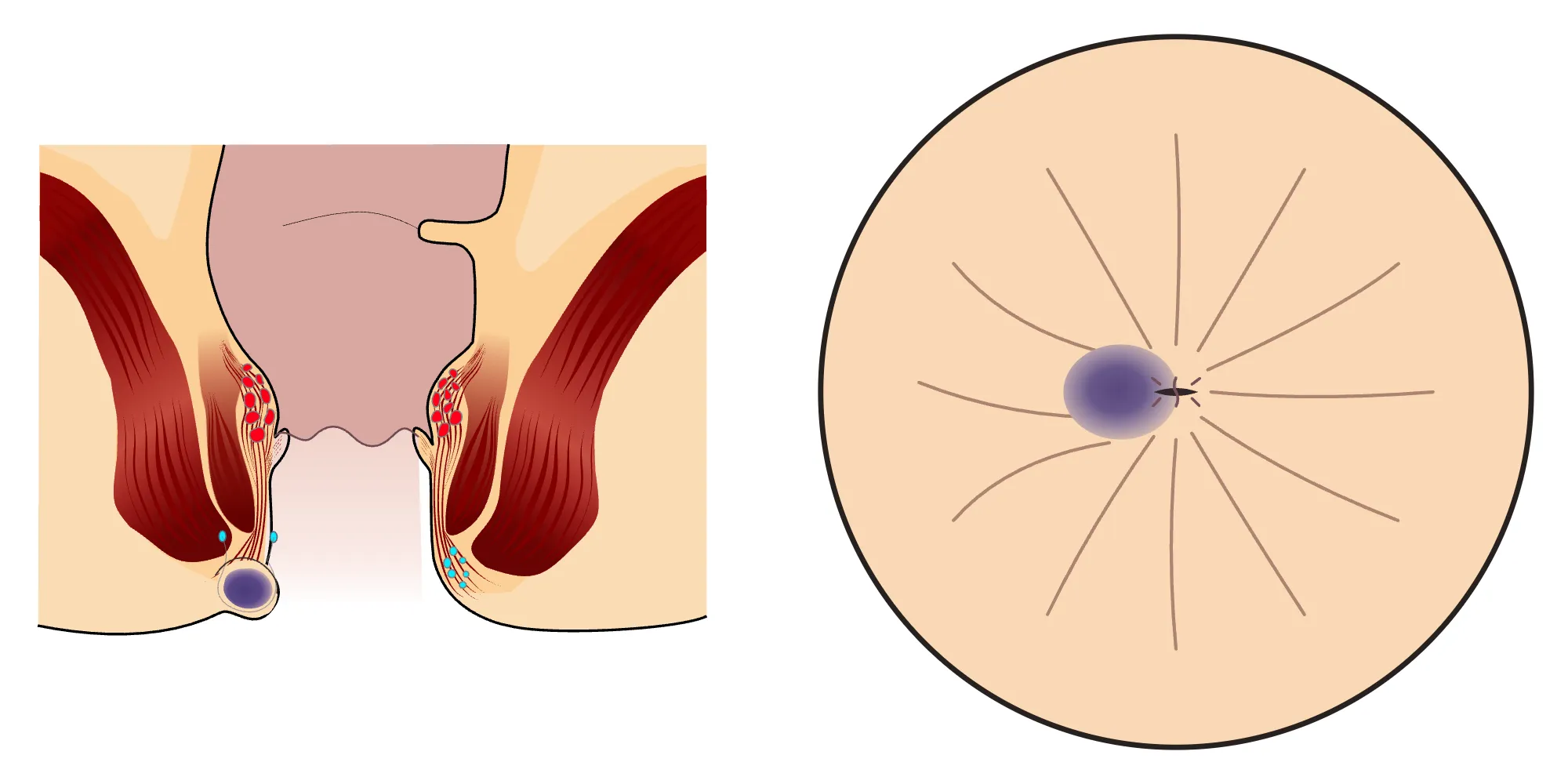
External view
- 1Clot
- 2Dentate line
- 3External view of the swelling caused by the clot
A painful, usually bluish swelling that appears suddenly at the edge of the anus may be caused by a clot in a blood vessel beneath the skin (perianal thrombosis or external haemorrhoid thrombosis); a doctor assesses the cause by examination and requests further tests if needed. If the swelling is red or warm to the touch, or if you have a fever, feel cold or have chills, there may be another cause, such as an abscess: see a doctor the same day. The clot in the drawing is beneath the skin, below the dentate line; from the outside it appears as a tense, bluish-purple swelling at the edge of the anus. The anal canal is drawn open to show the structures; it is normally closed.[4, 5, 8, 19]
Illustrative drawing, not to scale. Source: Armin Kübelbeck, CC BY 3.0. The source drawing “Analthrombose 01” was adapted. The colour inside the bowel was changed, the inner surface below the dentate line was coloured light, a clot together with the blood vessel wall was drawn inside the swelling, and a new external view was added on the right; numbered labels were added. The cross-sectional and external views were shown in separate panels.
Enlarge
Swipe the drawing right or left to see the details. You can use the arrow keys on your keyboard.
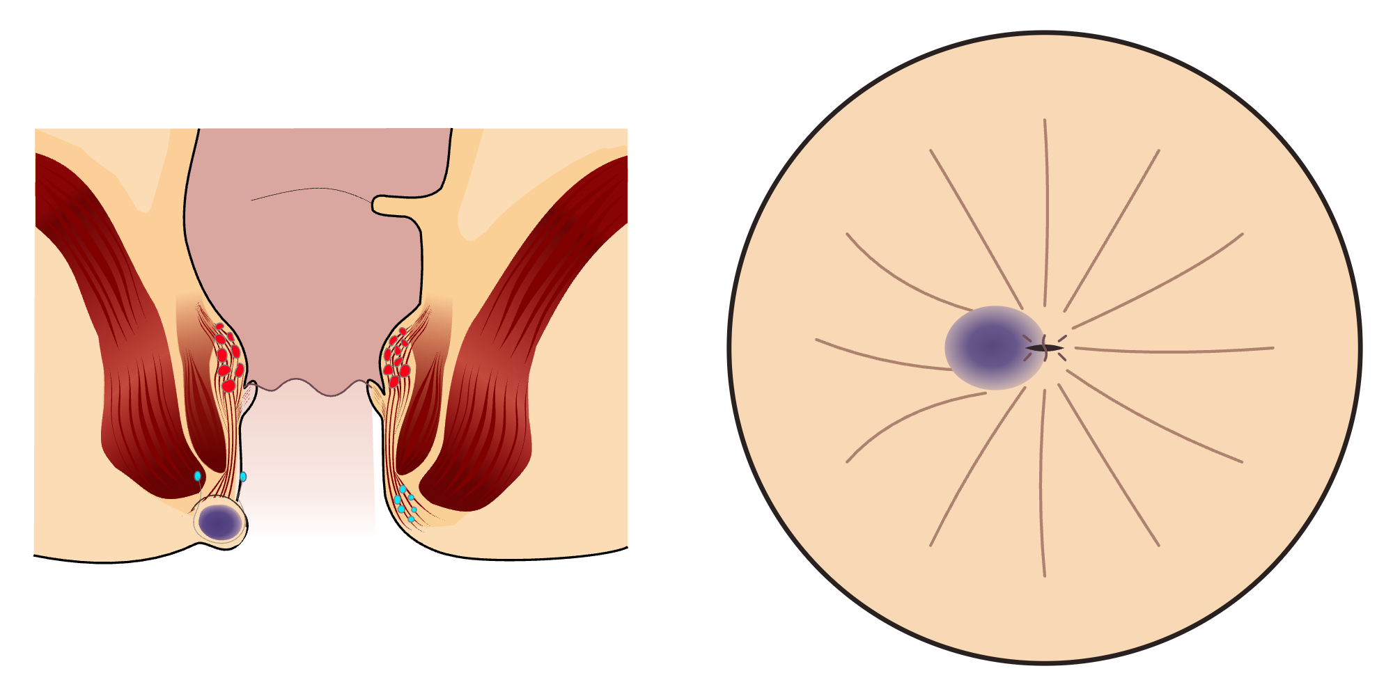
According to the Italian consensus statement, complete removal of the clotted tissue in a minor procedure reduces pain and recurrence more than simply cutting it and draining the clot or treatment with nitroglycerin cream; according to the statement, most patients benefit when this procedure is performed within the first 72 hours after symptoms begin[2]. The more recent American guideline states that early removal may be beneficial only in selected patients and grades this as a weak recommendation[5]. You and your doctor decide together whether to remove it, based on when the symptoms began and the examination findings. For internal haemorrhoids that prolapse, swell and develop a clot, treatment is usually initially conservative and with medicines; surgery is discussed for selected patients[1, 3]. Few studies have been conducted on this subject[1].
Haemorrhoids during pregnancy
Haemorrhoids are common during pregnancy, particularly in the last three months and in the first month after childbirth[2]. In most women, symptoms go away on their own shortly after childbirth; treatment therefore focuses on relieving symptoms through dietary and lifestyle changes[1, 2].
Data on the safety during pregnancy of medicines used for haemorrhoids are limited[2, 3]; increasing fibre and fluid intake, softening the stool and improving toilet habits are therefore emphasised[2]. The European guideline recommends basic treatment consisting of stool softeners, creams, medicines that strengthen blood vessels and painkillers during pregnancy and after childbirth[1]. The Italian statement says that medicines from the rutoside group, taken by mouth to strengthen blood vessels, cannot be recommended until new evidence on their safety during pregnancy becomes available[2]. According to the German guideline, medicines should be used very sparingly during pregnancy and breastfeeding, including for haemorrhoids, which usually follow a harmless course[3]; your doctor decides which medicine is suitable, so use medicines only after consulting your doctor.
After surgery
Controlling pain by combining more than one method supports recovery and helps reduce the risk of being unable to urinate[2, 5]. This is different from being unable to pass stool: in some patients, urine cannot be emptied after surgery even when the bladder is full (urinary retention)[5]. After surgery, the anal area is cleaned by washing it gently with clean water in the shower[3].
Recovery time varies depending on the method and the person. Studies have reported a return to work at about one to four weeks after classic surgery; with the classic method in which the wound is left open (Milligan–Morgan), the wound may take four to eight weeks to heal completely[3].
The rates below are results from different studies compiled by the Italian consensus statement for haemorrhoid operations; they are not the results of any individual doctor:
- Significant bleeding: on average around 2% after operations that remove haemorrhoidal tissue (the classic method or a procedure using a bipolar vessel-sealing device); the need for another procedure because of bleeding is 1–2%[2].
- Inability to urinate (urine remaining in the bladder): one of the most common problems; reported in studies at between 3% and 50%, and around 15% in most. It usually resolves on its own, but some patients need urine to be drained with a catheter; if you cannot urinate after surgery, contact your healthcare team without delay[2].
- Anal narrowing: between 1% and 7.5% after the stapled procedure or removal of haemorrhoidal tissue[2].
- Faecal incontinence: reported at approximately 6% after classic surgery to remove haemorrhoidal tissue and up to 5% after the stapled procedure[2].
- Recurrence of piles: between 2% and 8% after conventional surgery[2].
Although very rare, life-threatening complications have been reported after all types of procedures and surgery for piles; this possibility should be discussed before the procedure[2, 4].
Sources
23
- van Tol RR, Kleijnen J, Watson AJM, Jongen J, Altomare DF, Qvist N, et al. European Society of ColoProctology: guideline for haemorrhoidal disease. Colorectal Dis. 2020;22(6):650–662. doi: 10.1111/codi.14975. PubMed: 32067353.
- Gallo G, Martellucci J, Sturiale A, Clerico G, Milito G, Marino F, et al. Consensus statement of the Italian society of colorectal surgery (SICCR): management and treatment of hemorrhoidal disease. Tech Coloproctol. 2020;24(2):145–164. doi: 10.1007/s10151-020-02149-1. PubMed: 31993837.
- Deutsche Gesellschaft für Koloproktologie. S3-Leitlinie Hämorrhoidalleiden (AWMF-Register Nr. 081/007), 2019. Version marked as expired by AWMF. Text: register.awmf.org.
- van Tol RR, Kleijnen J, Watson AJM, et al. European Society of ColoProctology (ESCP) Guideline for Haemorrhoidal Disease, expanded version, 2019. Text: www.escp.eu.com.
- Hawkins AT, Davis BR, Bhama AR, Fang SH, Dawes AJ, Feingold DL, Lightner AL, Paquette IM. The American Society of Colon and Rectal Surgeons Clinical Practice Guidelines for the Management of Hemorrhoids. Dis Colon Rectum. 2024;67(5):614–623. (Full text reviewed: 1 October 2026.) doi: 10.1097/DCR.0000000000003276. PubMed: 38294832.
- NHS (United Kingdom National Health Service). Piles (haemorrhoids): patient information page. Last reviewed: 1 April 2026. Text: www.nhs.uk.
- Albuquerque A. Rubber band ligation of hemorrhoids: A guide for complications. World J Gastrointest Surg. 2016;8(9):614–620. doi: 10.4240/wjgs.v8.i9.614. PubMed: 27721924.
- NHS (United Kingdom National Health Service). Skin abscess (skin abscess): patient information page. Last reviewed: 2 June 2023. Text: www.nhs.uk.
- NHS (United Kingdom National Health Service). Anal pain (pain in the anus): patient information page. Last reviewed: 27 October 2025. Text: www.nhs.uk.
- American Gastroenterological Association. Hemorrhoids: Symptoms, causes, and treatments. Patient information; updated: May 2026. Text: patient.gastro.org.
- National Institute of Diabetes and Digestive and Kidney Diseases (NIDDK). Eating, Diet, & Nutrition for Hemorrhoids. Text: www.niddk.nih.gov.
- World Health Organization. WHO guidelines on physical activity and sedentary behaviour: at a glance. 2020. General adult health recommendations. Text: www.who.int.
- Brillantino A et al. The Italian Unitary Society of Colon-Proctology (SIUCP) guidelines for the management of acute and chronic hemorrhoidal disease. Ann Coloproctol. 2024;40(4):287–320. doi: 10.3393/ac.2023.00871.0124. Text: pmc.ncbi.nlm.nih.gov.
- Qureshi W, Hoang S, Frye J, Rao SSC. AGA Clinical Practice Update on Diagnosis and Treatment of Hemorrhoids: Expert Review. 2026. Official AGA recommendations reviewed; the full text on the publisher’s site could not be accessed. doi: 10.1016/j.cgh.2026.04.008. Text: gastro.org.
- Guy’s and St Thomas’ NHS Foundation Trust. Anal fissure treatment: Overview. Assessment under anaesthesia when an anal examination cannot be performed because of pain. Text: www.guysandstthomas.nhs.uk.
- Ambe PC and LHP Recommendation Development Group. Best clinical practice recommendations for the management of symptomatic hemorrhoids via laser hemorrhoidoplasty: the LHP recommendations. Tech Coloproctol. 2025;29:2. Published online: 23 November 2024. doi: 10.1007/s10151-024-03022-1. Text: pmc.ncbi.nlm.nih.gov.
- Ratto C. THD Doppler procedure for hemorrhoids: the surgical technique. Tech Coloproctol. 2014;18:291–298. Text: pmc.ncbi.nlm.nih.gov.
- National Institute of Diabetes and Digestive and Kidney Diseases (NIDDK). Colonoscopy. Official patient information; last reviewed: August 2023. Full page reviewed: 3 October 2026. Text: www.niddk.nih.gov.
- American Society of Colon and Rectal Surgeons (ASCRS). Hemorrhoids: Expanded Information. Official patient information; no visible update date is given. Full page reviewed: 3 October 2026. Text: www.fascrs.org.
- UCSF Department of Surgery. Hemorrhoidectomy. Image source: Andrea C. Bafford and Ronald Bleday, Hemorrhoids, Knol version 20, 28 July 2008. No visible update date is given on the page; full page reviewed: 4 October 2026. Text: staging16surgery.ucsf.edu.
- Ambe PC. Laserbehandlung beim Hämorrhoidalleiden. coloproctology. 2026;48:259–265. Published online: 27 April 2026. Figure 3 and full text reviewed: 4 October 2026. doi: 10.1007/s00053-026-00961-1. Text: link.springer.com.
- Wu X, Wen Q, Cui B, et al. Cap-assisted endoscopic sclerotherapy for internal hemorrhoids: technique protocol and study design for a multi-center randomized controlled trial. Ther Adv Gastrointest Endosc. 2020;13. Published online: 5 June 2020. Research protocol; Figure 3 and full text reviewed: 4 October 2026. doi: 10.1177/2631774520925636. Text: journals.sagepub.com.
- McLemore EC, Rai R, Siddiqui J, et al. Novel endoscopic delivery modality of infrared coagulation therapy for internal hemorrhoids. Surg Endosc. 2012;26:3082–3087. Published online: 31 May 2012. Full text reviewed: 4 November 2026. doi: 10.1007/s00464-012-2325-1. Text: link.springer.com.
Medical review of the Turkish original: Op. Dr. Ömer Seyfullah Hasıripi, . This page is a translation checked by AI reviewers; the Turkish version prevails.
This page is for general information; it does not replace an examination. See a doctor about your symptoms.