
What is an anal abscess and what causes it?
An anal abscess is an infection that develops in the soft tissue around the anus[2]. There are small glands that open onto the inner surface of the anal canal; extensions of some of these glands pass through the inner muscle that keeps the anus closed (internal sphincter muscle)[1]. An abscess is thought to begin when one of these glands becomes blocked and infected[1].
An abscess most often develops immediately at the edge of the anus or in the fatty tissue beside the anus; abscesses that develop between the sphincter muscles or deeper are less common[2]. Symptoms and treatment vary greatly according to the location of the abscess[2].
Most abscesses have no other underlying disease; some, however, may occur as part of another disease, most often Crohn’s disease (an inflammatory bowel disease)[2]. Anal abscesses are common; they occur most often between the ages of 20 and 40 and more often in men than in women[2].
An abscess is also linked to an anal fistula: a fistula is an abnormal tunnel that forms between the inside of the anal canal and the skin around the anus, and most often develops after an abscess[1].
What are the symptoms of an anal abscess?
An abscess usually appears as a painful, red swelling at the edge of the anus; pus may or may not come from the swelling[1]. The pain usually starts suddenly; fever is also common[2, 3]. Constant throbbing pain, pus and blood on the paper when wiping, and a high temperature may be signs of an abscess or fistula[5].
Deep abscesses are harder to notice[2]. With such an abscess, pain may spread to the perineum (the area between the anus and the genitals), lower back or buttocks, or it may resemble an illness arising inside the abdomen; sometimes there is an inability to pass urine[2]. In older and debilitated people, people with diabetes and those with a weakened immune system, symptoms are often mild or may even be absent; for this reason, greater care and speed are needed in these people[2].
Although rare, an infection (Fournier’s gangrene) can occur that spreads very quickly through the tissues under the skin of the perineum or genitals, causes tissue death and can be life-threatening; about one fifth of cases start in the anal and large bowel region[2]. The most common symptoms are pain, swelling and redness in the perineum or scrotum; the pain is usually very severe, even when little can be seen from the outside[2]. Fever and a fast pulse are also common[2]. Because a delay in treatment increases the risk of death, the guideline recommends surgery as early as possible[2].
How is it diagnosed?
Diagnosis is usually made by listening to your symptoms and examining the anus, including with a finger[2]. If the pain is very severe, the examination may be performed under sedation or anaesthesia[2]. Blood tests and imaging are often unnecessary for diagnosis; nevertheless, blood sugar tests are recommended to detect undiagnosed diabetes[2]. Imaging (MRI, computed tomography or ultrasound through the anus) is recommended if symptoms are unusual or a deep abscess, complex fistula or Crohn’s disease is suspected[2].
Which department treats an anal abscess?
The general surgery department treats anal abscesses. Your family doctor or workplace doctor may perform the initial examination and, if they consider it necessary, refer you to general surgery. However, if you have a painful swelling or a fever, see a doctor the same day without waiting for an appointment[2, 4].
How is an anal abscess treated?
In adults, treatment of an anal abscess involves surgically opening the abscess and draining the pus (drainage)[2, 3]. Do not try to open or squeeze the abscess yourself: the infection may spread or the abscess may worsen[8]. If the abscess opens on its own, wash the area with soapy water, keep it clean and cover it, and still see a doctor: the infection may continue[8]. If there is a severe response of the body to infection (sepsis), diabetes, a weakened immune system or widespread inflammation of the skin, the abscess is drained urgently; if these are absent, it is ideally drained within 24 hours[2]. It is important to drain the abscess completely: in a study of simple anal abscesses, the abscess recurred in 41% of those whose pus was drawn out only with a needle, compared with 15% of those whose abscess was drained through an incision[2].
Are antibiotics needed?
After drainage, giving antibiotics to everyone to prevent a fistula is not recommended[1]. There is no meaningful evidence that antibiotics prevent fistulas; unnecessary use may also contribute to germs becoming resistant to medicines[1]. In most patients, antibiotics do not shorten the duration of treatment or reduce recurrence of the abscess[3]. Antibiotics are considered if there is sepsis, spreading inflammation of the skin around the abscess (cellulitis) or a weakened immune system (for example, due to chemotherapy, cancer or HIV), or if symptoms do not go away after drainage[1, 2, 3].
How is surgery for an anal abscess performed?
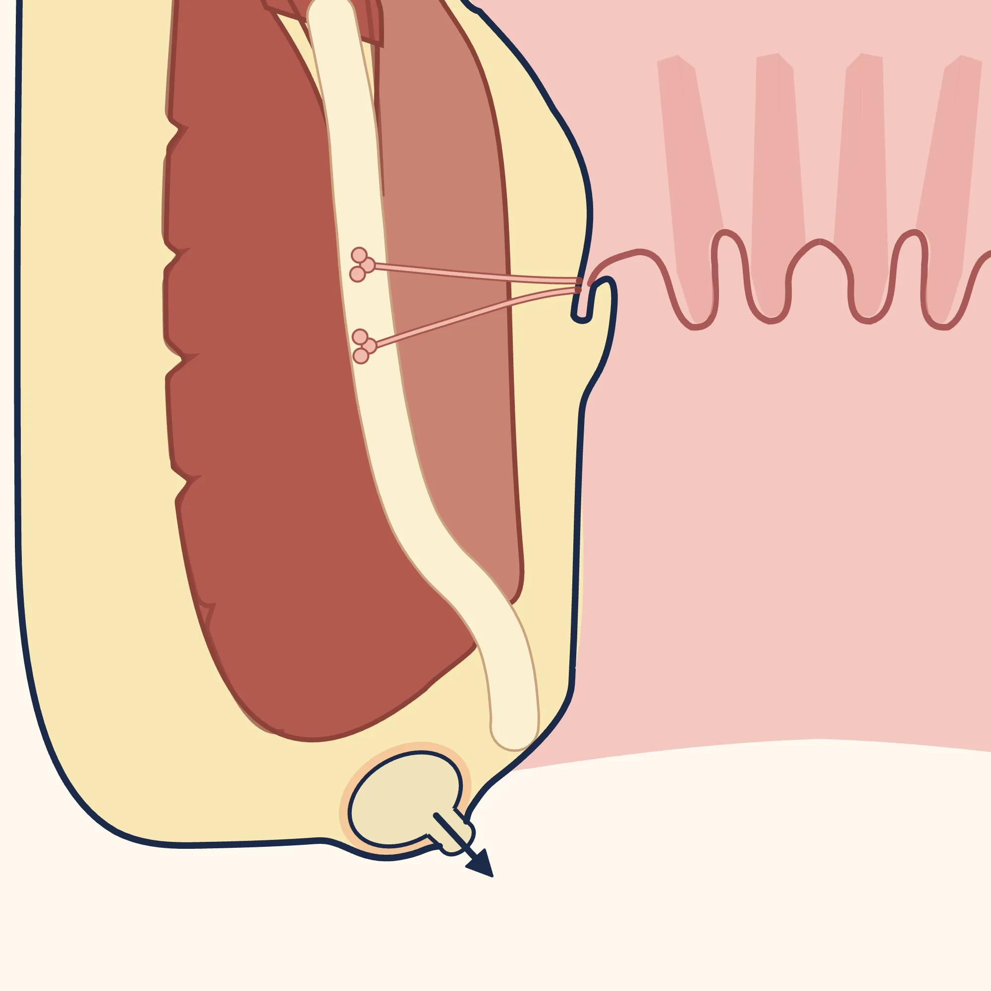
The position and size of the incision are determined by the type and location of the abscess[3]. As a general rule, the incision is made as close to the edge of the anus as possible, in a way that completely drains the abscess without damaging the muscles that keep the anus closed; the aim is to keep any fistula that develops later short[2, 3].
Enlarge
Swipe the drawing right or left to see the details. You can use the arrow keys on your keyboard.
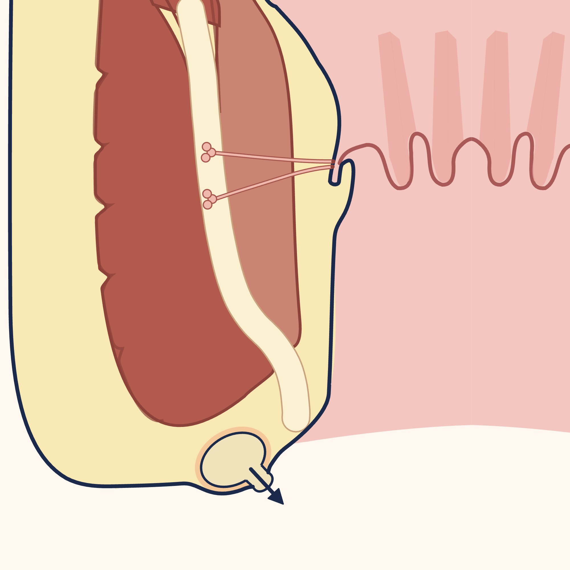
- 1 Anal canal
- 2 Edge of the anus
- 3 Internal sphincter muscle
- 4 External sphincter muscle
- 5 Abscess at the edge of the anus
- 6 Incision: close to the edge of the anus
- 7 Emptying the pus (drainage)
Description of the drawing
An abscess at the edge of the anus, as in the drawing, is drained through an incision in the skin over it (drainage). The incision is made as close to the edge of the anus as possible, in a way that completely drains the abscess without damaging the muscles that keep the anus closed; the aim is to keep any fistula that develops later short. The anal canal is shown open to display the structures; it is normally closed.[2, 3]
Results may vary from person to person with any surgical or interventional procedure. You are advised to discuss the procedure in detail with your doctor beforehand.
Illustrative drawing, not to scale. Source: Armin Kübelbeck, McortNGHH and Jmarchn, CC BY-SA 4.0. Adapted from Jmarchn’s “Abscess diag 02-es” drawing (CC BY-SA 3.0); that drawing is based on McortNGHH’s “Abscess diag 02” (CC BY-SA 4.0) and Armin Kübelbeck’s “Piles diffdiag 01” (CC BY 3.0). The text, arrows and abscess markings in the source were removed; the right half was mirrored to create a base with identical sides, the side outline of the body was redrawn, the colours were changed, the strip between the two muscles was widened, and anal glands opening into the canal at the level of the dentate line were added; the left wall and anal opening were shown close up; the fatty fringe at the anal opening was covered to sharpen the edge of the anus; an abscess pocket at the edge of the anus, a short skin incision above it, pus emerging from the incision and an arrow were added; numbered labels were added. This adaptation is also licensed under CC BY-SA 4.0.
In people who are otherwise healthy, do not have a weakened immune system and show no signs of sepsis, a small, superficial abscess may be drained by numbing only the area (local anaesthesia), without a hospital stay[2, 3]. For deep abscesses, numbing the area is not enough; in this situation, the Japanese guideline prefers anaesthesia given through the lower back or tailbone area (spinal or sacral epidural anaesthesia)[3].
The abscess still needs to be drained if you take a blood-thinning medicine[3]. Tell your doctor about your medicine: the method of anaesthesia is chosen accordingly[3]. Do not stop the medicine on your own; ask the doctor who prescribed it what to do. Antibiotics may be needed before drainage in situations such as a prosthetic heart valve, a previous infection of the inner lining of the heart (endocarditis), or certain congenital heart conditions; tell your doctor if you have such a condition[2].
Is the wound packed?
The European Society of Coloproctology (2024) recommends against routinely packing the wound with gauze after drainage: packing does not prevent a fistula or the abscess from coming back, but it increases pain[1]. Packing may be useful when bleeding needs to be stopped[1]. The older World Society of Emergency Surgery guideline (2021) stated that, based on the available studies, no recommendation could be made on this[2].
If a fistula is found during surgery
A fistula may be seen while the abscess is being drained, or it may show itself later through discharge or swelling[1]. The European Society of Coloproctology does not recommend cutting open the fistula during the same operation as the abscess drainage[1]. In studies, opening it at the same time reduced the chance of a fistula remaining afterwards (30% in those who had drainage alone, 5% in those whose fistula was opened at the same time); but at least half of the fistulas seen during drainage close on their own, and leakage of wind or stool one year later was three times more common in those whose fistula was opened at the same time[1]. The World Society of Emergency Surgery recommends opening a fistula at the same time only if it is clearly visible, lies just under the skin and does not involve the sphincter muscle; for a fistula involving the muscle, it recommends passing a loose thread (seton) through the tunnel[2]. For this reason, most often only the abscess is drained first; if a fistula remains, a separate operation may be needed later[1, 3].
What happens after surgery for an anal abscess?
An abscess may come back after it has been drained[2]. Inadequate drainage, compartments within the abscess, an abscess extending in a horseshoe shape, and a longer time between the start of symptoms and drainage increase this risk[2].
A fistula does not develop after an abscess in every patient; reported rates range from 15% to 37%[1, 2, 3]. A fistula increases the chance of the abscess coming back[2].
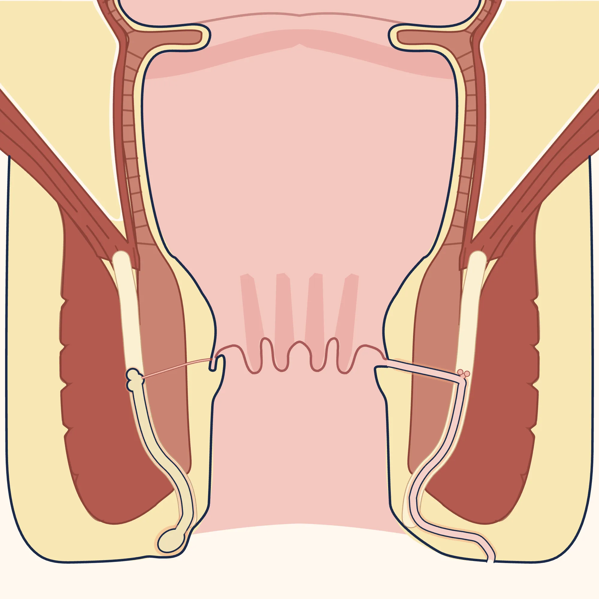
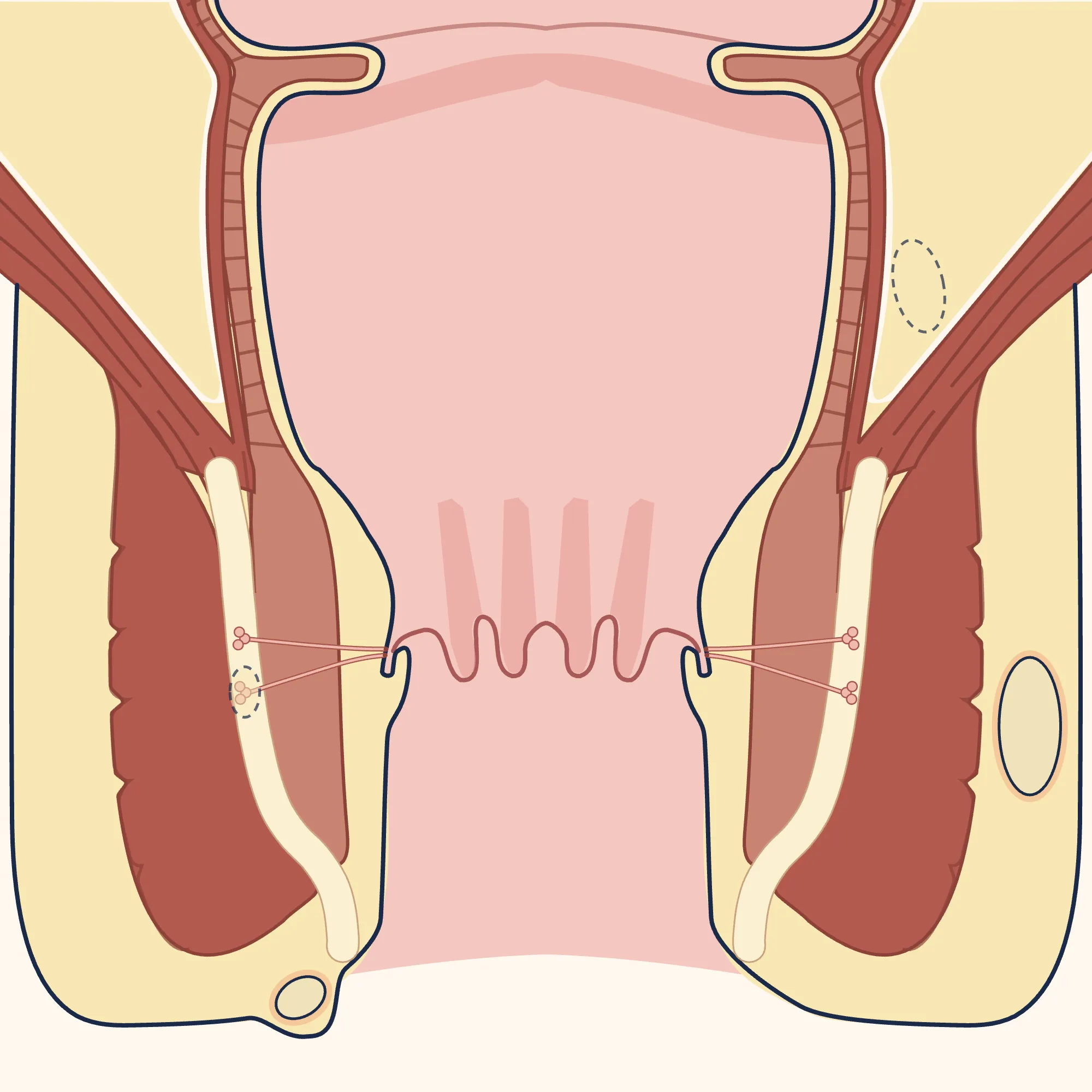
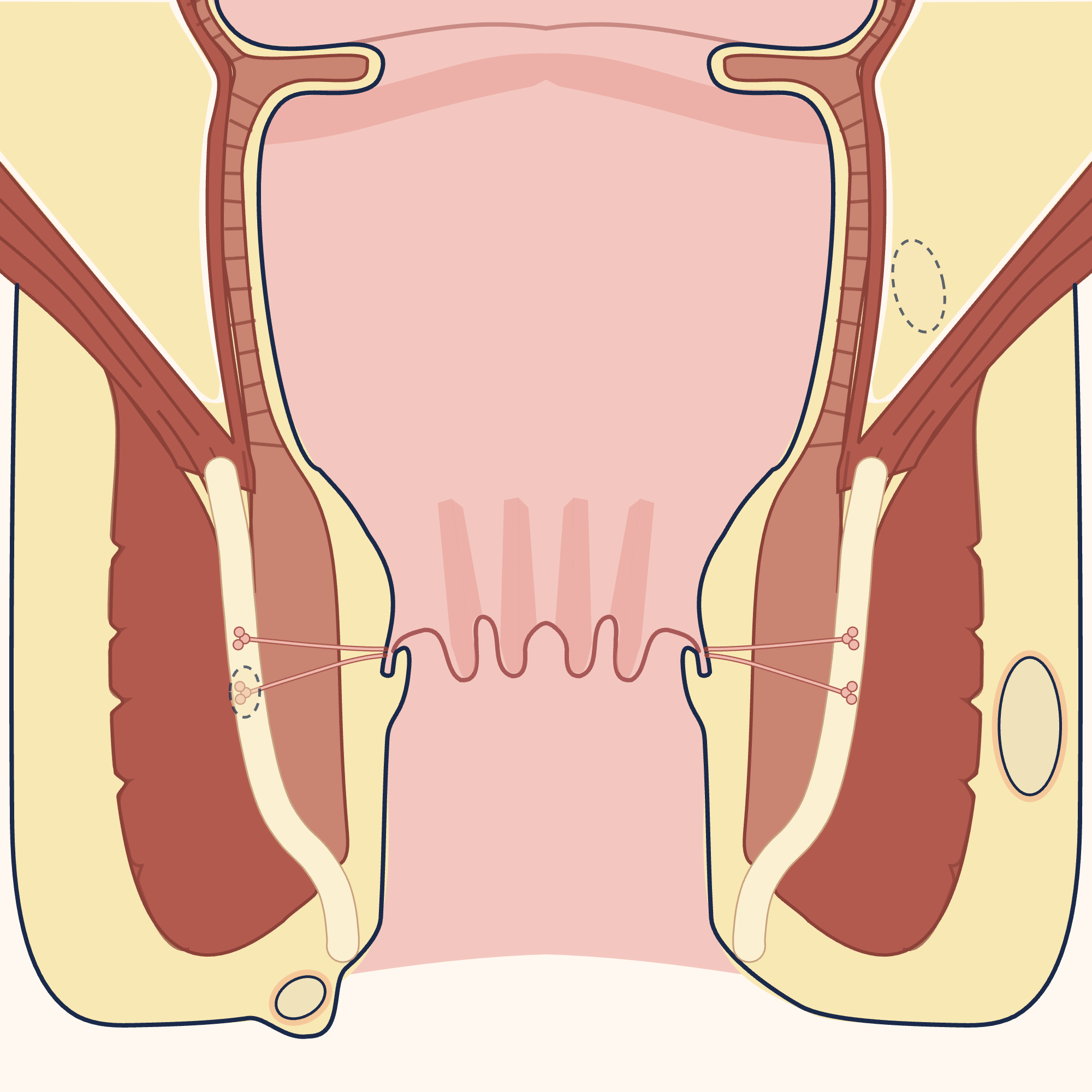
Enlarge
Swipe the drawing right or left to see the details. You can use the arrow keys on your keyboard.
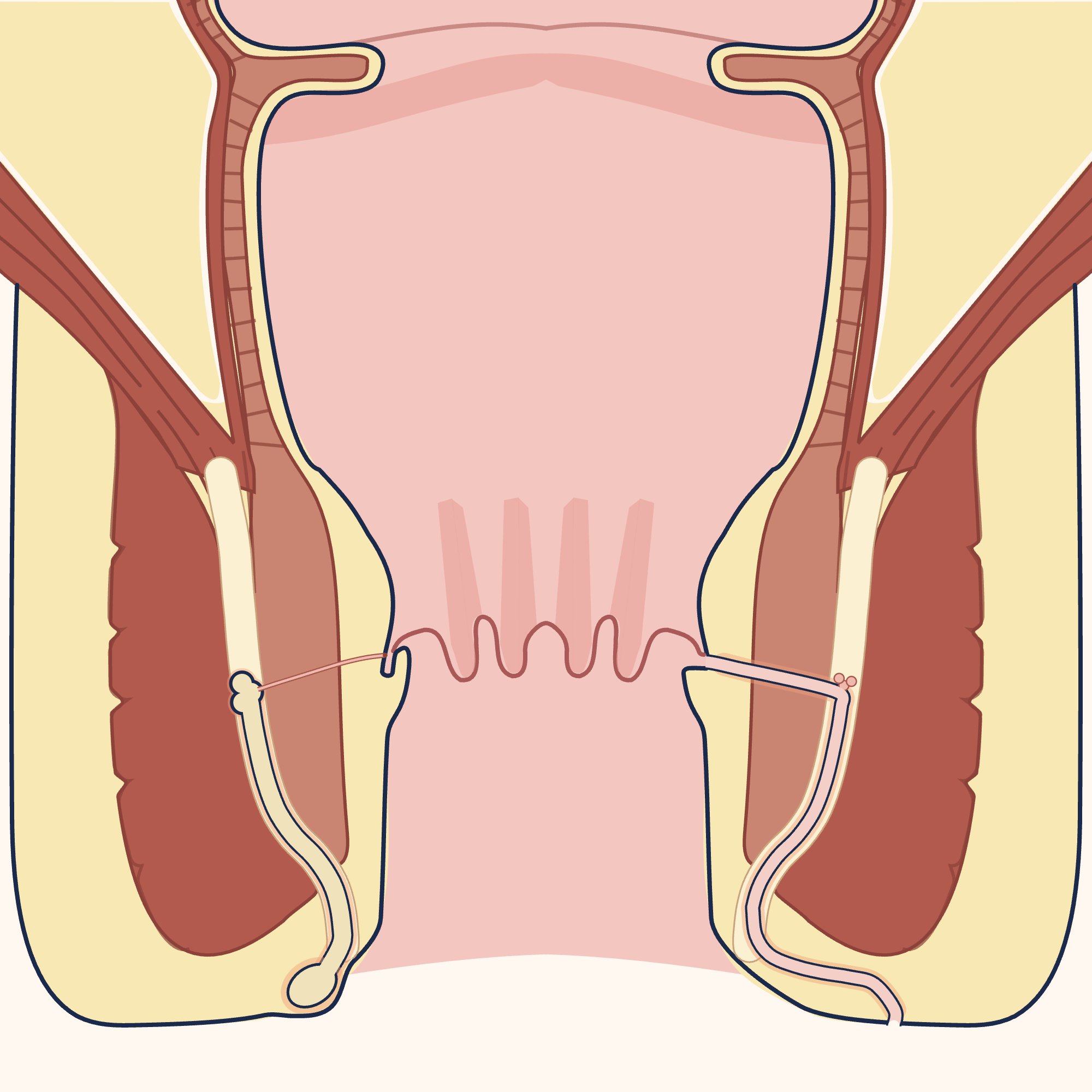
- 1 Gland opening onto the inner surface of the anus
- 2 Internal sphincter muscle
- 3 External sphincter muscle
- 4 Before: abscess starting in a gland
- 5 Internal opening
- 6 After: fistula tunnel
- 7 External opening (hole in the skin)
Description of the drawing
Left (before): an abscess that started in an anal gland has travelled down between the two muscles and collected at the edge of the anus. Right (after): the tunnel (fistula) that can remain after an abscess; its internal opening is where the gland opens into the canal, and its external opening is in the skin beside the anus. The drawing shows one of the possible paths of a fistula. The anal canal is shown open to display the structures; it is normally closed.[1, 3]
Illustrative drawing, not to scale. Source: Armin Kübelbeck, McortNGHH and Jmarchn, CC BY-SA 4.0. Adapted from Jmarchn’s “Abscess diag 02-es” drawing (CC BY-SA 3.0); that drawing is based on McortNGHH’s “Abscess diag 02” (CC BY-SA 4.0) and Armin Kübelbeck’s “Piles diffdiag 01” (CC BY 3.0). The text, arrows and abscess markings in the source were removed; the right half was mirrored to create a base with identical sides, the side outline of the body was redrawn, the colours were changed, the strip between the two muscles was widened, and anal glands opening into the canal at the level of the dentate line, the spread of an abscess on one side and a fistula tunnel on the other were added; numbered labels were added. This adaptation is also licensed under CC BY-SA 4.0.
With every abscess, particularly if it comes back, it is necessary to make sure there is no undiagnosed Crohn’s disease[2]. According to hospital records in the United Kingdom, 3% of people who had an anal abscess for the first time were later diagnosed with Crohn’s disease[1].
Routine imaging is usually not needed after drainage; imaging is recommended if the abscess comes back, if there is a fistula or a wound that does not heal, or if an inflammatory bowel disease is suspected[2]. See your doctor if the abscess comes back, if discharge or swelling around the anus continues, or if the wound does not heal[1, 2].
Sources
8
- Reza L, Gottgens K, Kleijnen J, Breukink S, Ambe PC, Aigner F, et al. European Society of Coloproctology: Guidelines for diagnosis and treatment of cryptoglandular anal fistula. Colorectal Dis. 2024;26(1):145–196. doi: 10.1111/codi.16741. PubMed: 38050857.
- Tarasconi A, Perrone G, Davies J, Coimbra R, Moore E, Azzaroli F, et al. Anorectal emergencies: WSES-AAST guidelines. World J Emerg Surg. 2021;16(1):48. doi: 10.1186/s13017-021-00384-x. PubMed: 34530908.
- Yamana T. Japanese Practice Guidelines for Anal Disorders II. Anal fistula. J Anus Rectum Colon. 2018;2(3):103–109. doi: 10.23922/jarc.2018-009. PubMed: 31559351.
- NHS (United Kingdom National Health Service). Skin abscess (skin abscess): patient information page. Last reviewed: 2 June 2023. Text: www.nhs.uk.
- NHS (United Kingdom National Health Service). Anal pain (pain in the anus): patient information page. Last reviewed: 27 October 2025. Text: www.nhs.uk.
- NHS (United Kingdom National Health Service). Sepsis: patient information page. Last reviewed: 14 May 2026. Text: www.nhs.uk.
- healthdirect Australia (Australian national health information service). Urinary retention (urinary retention): patient information page. Last reviewed: July 2024. Text: www.healthdirect.gov.au.
- Cleveland Clinic. Perianal Abscess (abscess around the anus): patient information page. Last updated: 17 June 2022. Text: my.clevelandclinic.org.
Medical review of the Turkish original: Op. Dr. Ömer Seyfullah Hasıripi, . This page is a translation checked by AI reviewers; the Turkish version prevails.
This page is for general information; it does not replace an examination. See a doctor about your symptoms.