
What is an anal fistula and what causes it?
An anal fistula is an abnormal tunnel that forms between the inside of the anus and the skin around it[1]. It usually develops after an anal abscess (a collection of pus around the anus)[1].
Abscesses and fistulas are thought to usually start when one of the glands opening onto the inner surface of the anus becomes blocked and inflamed[1, 2]. As the continuing inflammation finds the easiest way out, it usually forms a path through or between the muscles that keep the anus closed (sphincter muscles)[1]. A fistula may already be present when the abscess is drained (at least half of these close on their own), or it may appear later[1].
What are the symptoms of an anal fistula?
The most common symptoms are discharge containing pus, or swelling and pain around the anus that come and go[2]. The discharge comes from an opening in the skin around the anus; as well as pus, mucus, blood or watery stool may come out[1]. A fistula can also cause discomfort and skin irritation[6]. If there is a red swelling around the anus and a fever, an abscess may also have developed alongside the fistula[6].
How is an anal fistula diagnosed?
The doctor listens to your symptoms and examines the anal region; during this examination, they also look for other causes that could lead to a fistula[1]. If your bowel habits, family history of illness or examination findings suggest Crohn’s disease (a type of inflammatory bowel disease), the inside of the bowel should also be examined[1].
MRI or ultrasound performed through the anus (endoanal ultrasound) should be readily used to distinguish between a simple and a complex fistula[1]. Sources differ on which to start with: the European radiology society ESGAR (2020) recommends MRI as the main imaging method[3]; the European Society of Coloproctology (2024) recommends MRI for a fistula that appears complex and says ultrasound may be considered as the first examination in most patients[1].
Which department treats an anal fistula?
The general surgery department treats anal fistulas. Your family doctor or workplace doctor may carry out the initial examination and, if they consider it necessary, refer you to general surgery.
How does an anal fistula go away and what helps?
An established fistula rarely closes on its own in adults[2]. Surgery is therefore the mainstay of treatment in adults[2, 6]; the German guideline (2026) recommends against non-surgical treatment for a fistula that starts in a gland[5]. If a fistula is due to a disease such as Crohn’s disease or tuberculosis, that disease also needs to be treated[2]. An untreated fistula can lead to repeated abscesses and, when it persists for a very long time, rarely to cancer[2].
Smoking, diabetes and excess weight can impair wound healing; the European guideline calls for these to be discussed with you before repair surgery[1]. In a study using a tissue flap, the fistula healed in 79% of non-smokers and 60% of smokers; however, there is no strong evidence showing the effect of these factors on fistula healing[1].
Is fistula surgery difficult?
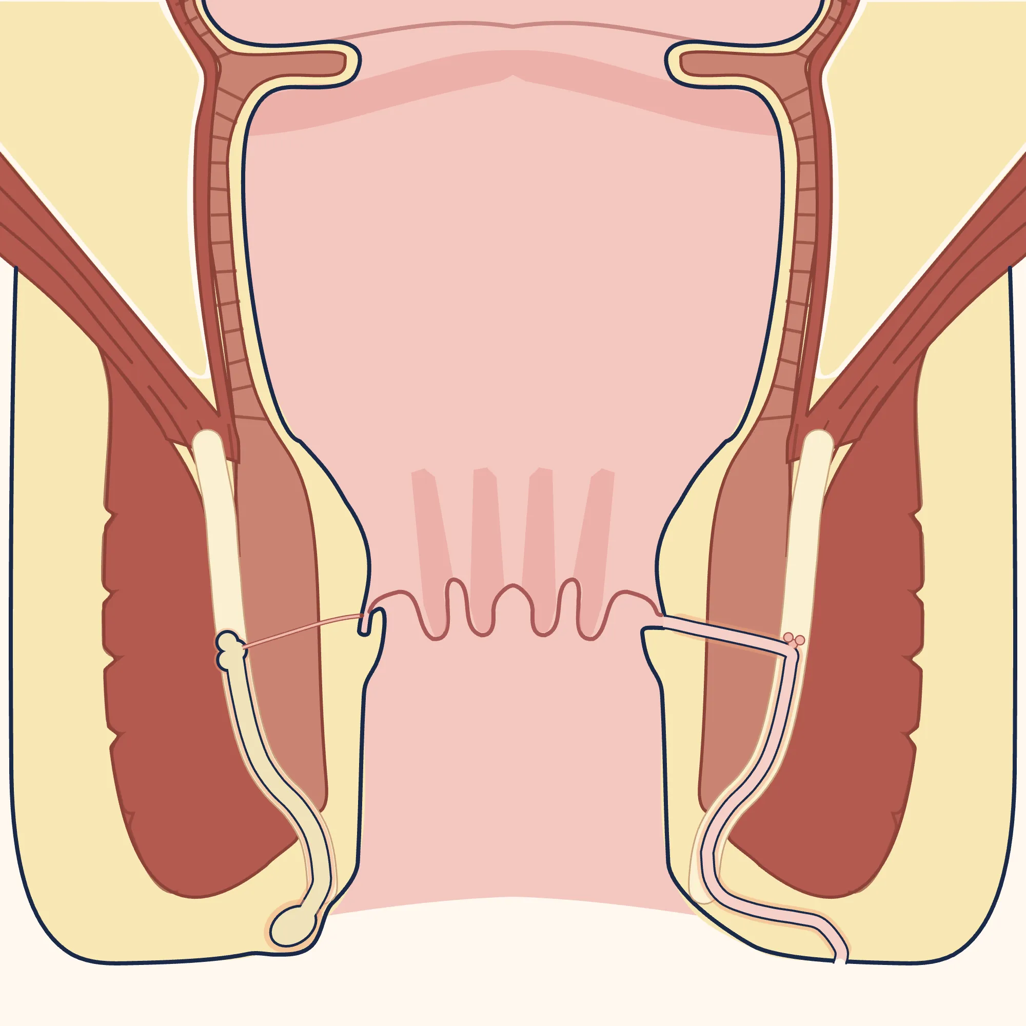
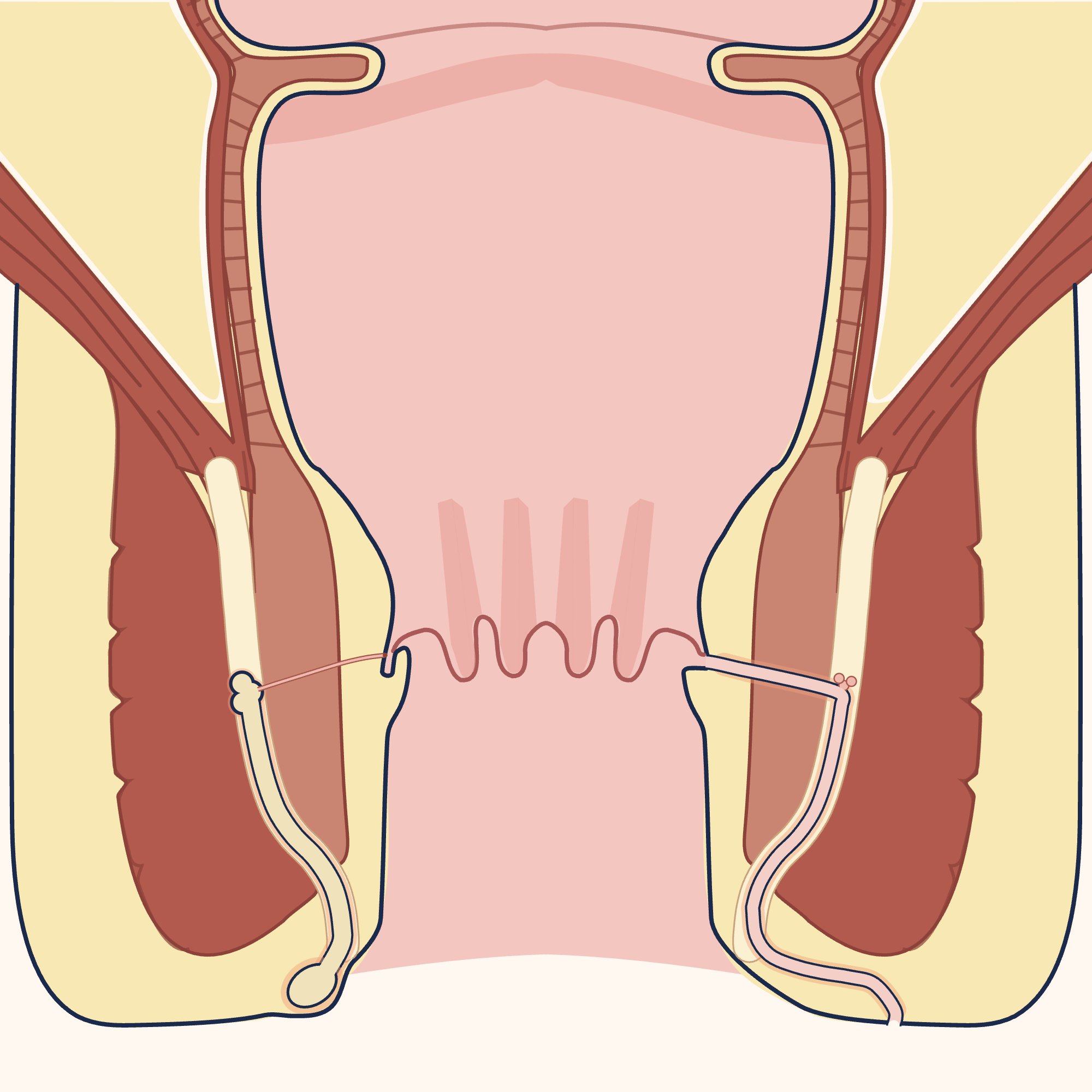
This depends on the type of fistula: fistulas are classified according to their relationship to the internal and external muscle layers that keep the anus closed[1, 2]. Fistulas passing beneath the skin, between the two muscle layers or through a small lower part of the external muscle are usually considered simple[1]. Fistulas that pass through more than 30% of the external muscle or more than half of the internal muscle, or loop over or outside the muscles, are considered complex; so are fistulas that recur, are at the front of the anus in women, are horseshoe-shaped or have multiple branches[1]. Fistulas passing through the upper part of the muscles are called “high fistulas”; however, there is no universally accepted definition of this[1].
Enlarge
Swipe the drawing right or left to see the details. You can use the arrow keys on your keyboard.
- 1 Internal sphincter muscle
- 2 External sphincter muscle
- 3 Low, passing between the two muscle layers (simple)
- 4 Looping over the muscles (complex)
- 5 Looping outside the muscles (complex)
- 6 Passing beneath the skin (simple)
- 7 Passing through the lower part of the external muscle (simple)
- 8 Passing through the upper part of the external muscle (complex)
Description of the drawing
Fistulas are classified according to their relationship to the two muscle layers that keep the anus closed. Left: low tunnels passing between the two muscles (simple), and tunnels looping over and outside the muscles (complex). The type that loops outside the muscles is rare and usually has another cause. Right: tunnels passing beneath the skin (simple), through the lower part of the external muscle (simple), and through the upper part of the external muscle (complex). Six tunnels are shown in one drawing for comparison. The anal canal is shown open to display the structures; it is normally closed.[1, 5]
Illustrative drawing, not to scale. Source: Armin Kübelbeck, McortNGHH and Jmarchn, CC BY-SA 4.0. Adapted from Jmarchn’s “Abscess diag 02-es” drawing (CC BY-SA 3.0); that drawing is based on McortNGHH’s “Abscess diag 02” (CC BY-SA 4.0) and Armin Kübelbeck’s “Piles diffdiag 01” (CC BY 3.0). The text, arrows and abscess markings in the source were removed; the right half was mirrored to create a base with identical sides, the side outline of the body was redrawn, the colours were changed, and the strip between the two muscles was widened (its lower end was shortened and, on the left wall, it was extended to the fatty space above the pelvic floor muscle); three fistula tunnels with their internal and external openings were added to each wall; numbered labels were added. This adaptation is also licensed under CC BY-SA 4.0.
A simple fistula may be suitable for opening the tunnel; a complex fistula may not close despite several attempts at repair, and the risk of damaging the muscles is higher[1].
How is fistula surgery performed?
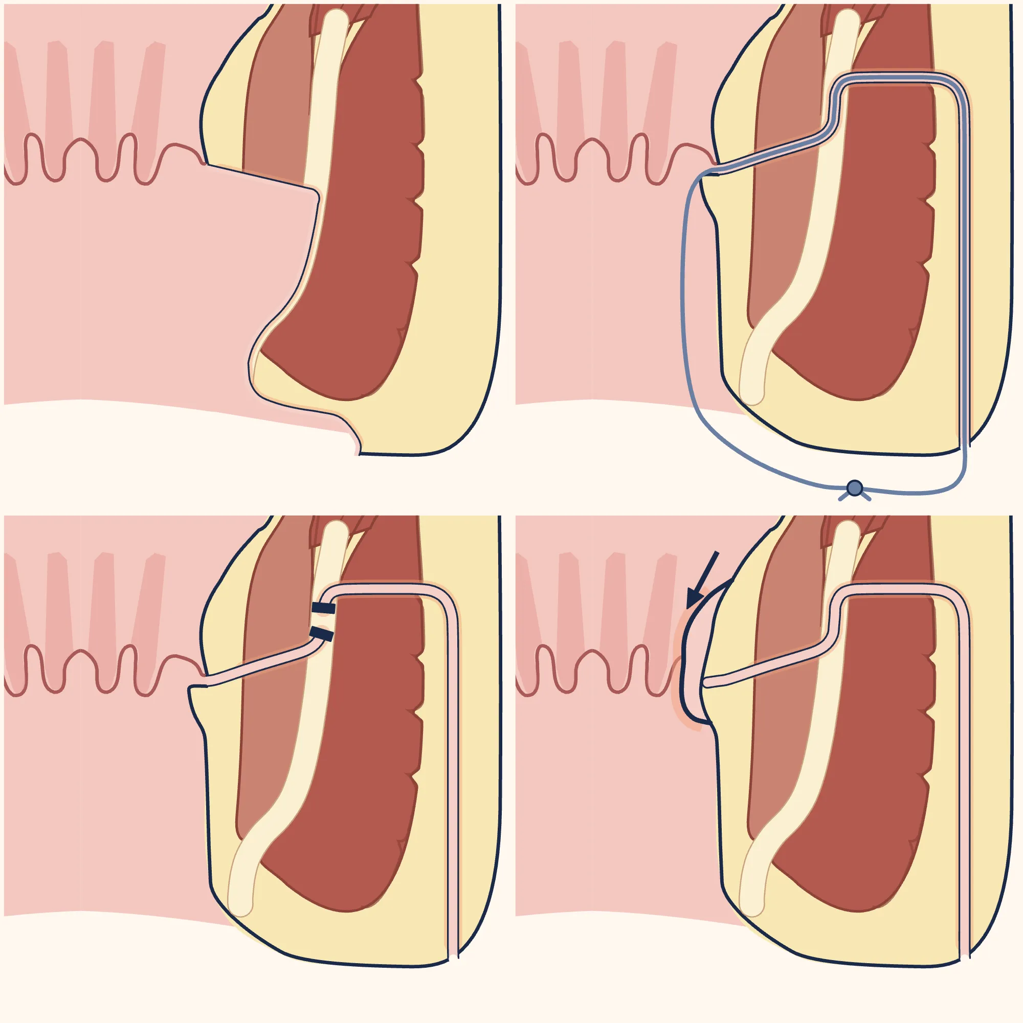
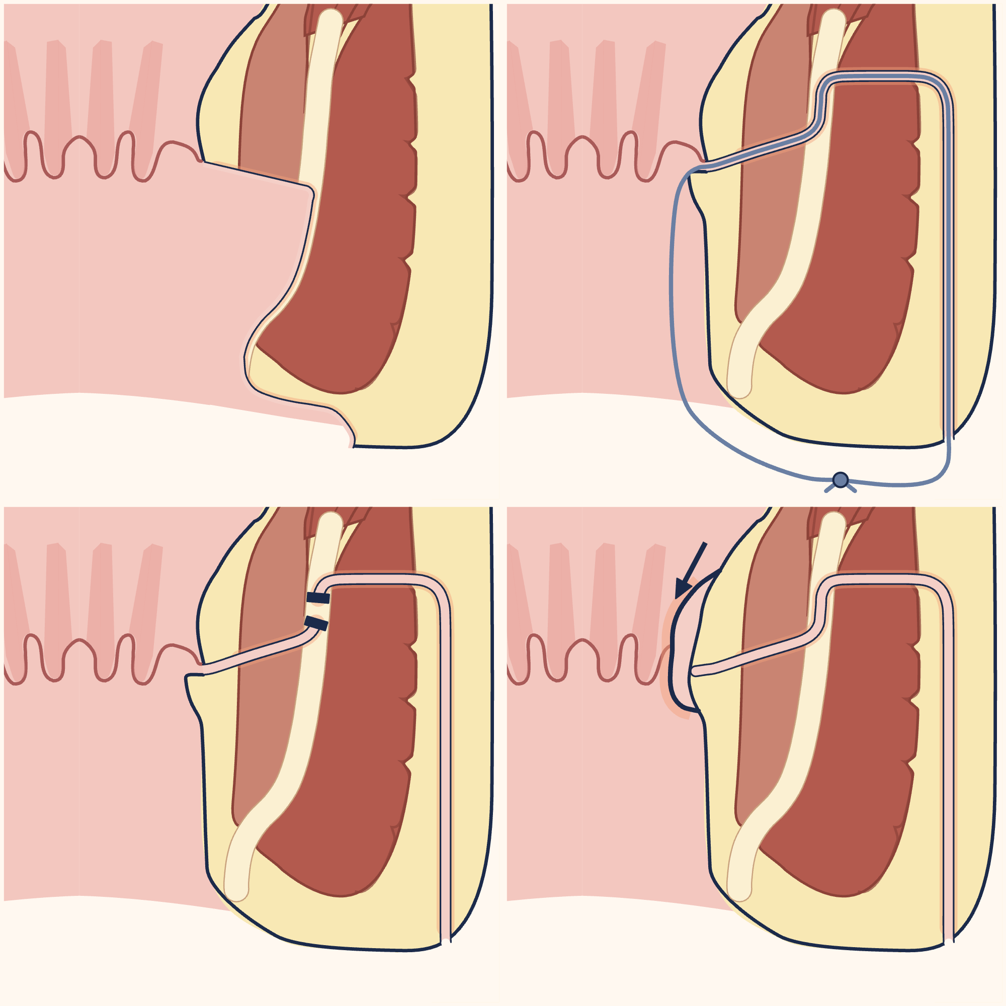
After an examination and, if needed, an MRI or ultrasound, the operation is chosen together with your surgeon according to the type of fistula, the function of your anal muscles, your expectations and how much risk of impaired bowel control you are willing to accept; more than one method may be suitable for the same situation[1, 2].
The essence of the choice is this: in simple fistulas, the fistula healed in an average of 93.7% with methods that open the tunnel by also cutting the muscle, and in 77.7% with muscle-preserving methods. On the other hand, after methods that cut the muscle, mild or severe leakage of stool was reported in 12.7% of patients and severe leakage in 6%; however, follow-up was shorter than one year in most of these studies[1]. This risk increases as the thickness of muscle cut increases[2, 5]. The rates on this page have been compiled from different studies and vary from study to study[1, 2]; the European guideline’s recommendations are mostly based on low or very low levels of evidence[1]. These rates are not any individual doctor’s own results.
Enlarge
Swipe the drawing right or left to see the details. You can use the arrow keys on your keyboard.
- 1 Opening the tunnel (fistulotomy): muscle in its path is also cut
- 2 Loose thread (drainage seton): does not tighten around the muscle
- 3 Tying the tunnel between the muscles (LIFT)
- 4 Tissue flap (advancement flap): muscle-preserving repair
- 5 Internal sphincter muscle
- 6 External sphincter muscle
- 7 Fistula tunnel
- 8 Internal opening
Description of the drawing
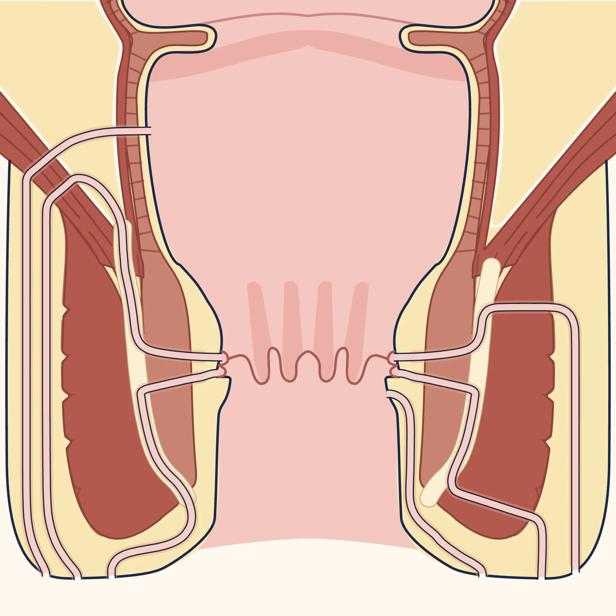
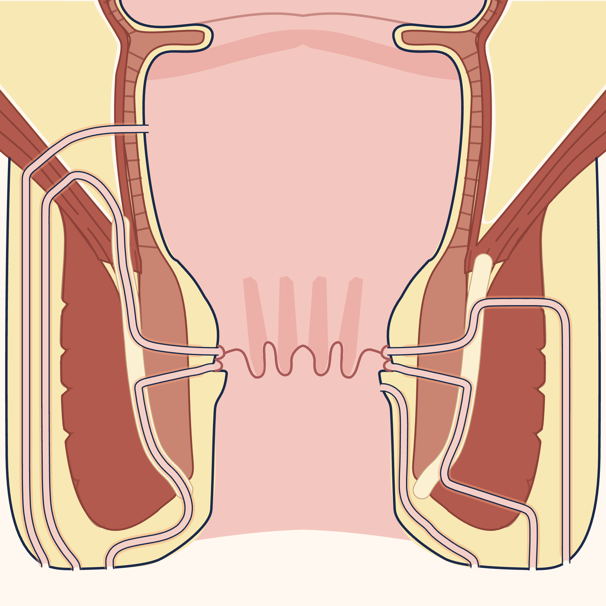
The main fistula operations are shown in four close-up drawings of the same cross-section of the anal region (from left to right, top to bottom): opening the tunnel along its entire length (for simple tunnels; any muscle in its path is also cut), passing a loose thread through the tunnel (the muscle is not tightened), tying and cutting the tunnel between the two muscles (LIFT), and covering the internal opening with a tissue flap taken from the inner wall of the bowel (the muscle is preserved). The anal canal is drawn open to show the structures; it is normally closed.[1, 7]
Results may vary from person to person with any surgical or interventional procedure. You are advised to discuss the procedure in detail with your doctor beforehand.
Illustrative drawing, not to scale. Source: Armin Kübelbeck, McortNGHH and Jmarchn, CC BY-SA 4.0. Adapted from Jmarchn’s “Abscess diag 02-es” drawing (CC BY-SA 3.0); that drawing is based on McortNGHH’s “Abscess diag 02” (CC BY-SA 4.0) and Armin Kübelbeck’s “Piles diffdiag 01” (CC BY 3.0). The text, arrows, abscess markings and glands in the source were removed; the right half was mirrored to create a base with identical sides, the side outline of the body was redrawn, the colours were changed and the strip between the two muscles was widened; the base was zoomed in on the right wall and repeated in four panels; a fistula tunnel was added to each panel, along with, respectively, the open groove of the tunnel, a loose thread and knot, two ties and a cut gap, and a tissue flap and arrow; numbered labels were added. This adaptation is also licensed under CC BY-SA 4.0.
Main methods
- Opening the tunnel (fistulotomy): The tunnel is opened from its internal opening to its external opening, and any muscle in its path is also cut[1]. After a detailed assessment, the European guideline recommends this for people whose fistula passes between the two muscle layers or through the lower part of the external muscle and who have normal bowel control[1]. Healing rates of 96–98% have been reported for these fistulas, and 83.6% in a large series followed for approximately 3 years[1]. Greater caution is needed in women with a fistula at the front of the anus or whose anus was injured during childbirth, and in people who have previously had anal surgery[1].
- Loose thread (drainage seton): A loose thread placed in the tunnel allows inflammatory material to drain and prepares the tunnel for an operation that preserves the muscle; it does not tighten the muscle[1]. Strong evidence has not shown that it closes the fistula on its own[1]. For a complex and recurrent fistula, it may be considered as a long-term solution if another procedure could impair bowel control or if you do not want another operation[1].
- Thread that is tightened (cutting seton): A thread tightened over time gradually cuts through and opens the tunnel[2]. The European (2024) and German (2026) guidelines recommend against its use[1, 5]; the Japanese guideline, based on studies up to 2013, finds it effective for low fistulas[2]. In one study, bowel control was impaired in 24% of patients; in another, 8.5% experienced severe faecal incontinence. Frequent check-ups are needed to tighten the thread[1].
- Tissue flap (advancement flap): This is a muscle-preserving repair in which the internal opening of the tunnel is closed with a piece of tissue prepared from the inner wall of the bowel[1, 5]. The European guideline recommends it for high fistulas[1]. Healing was reported at 74.6%, and recurrence at 21% over an average follow-up of two and a half years[1]. Faecal incontinence rose from 9.3% to 20.4% as the flap became thicker; most cases were mild[1].
- Tying the tunnel between the muscles (LIFT): The tunnel is located between the two muscle layers, then tied and cut there[1]. The European guideline recommends it for a fistula passing high through the external muscle that is being repaired for the first time[1]. Success was reported at 69.1% and faecal incontinence at 1.6%[1]. In two studies, healing was 85% with LIFT and 79.5% with a tissue flap; the difference was not significant, while pain was significantly lower with LIFT[1].
- Laser (LAFT) and camera-assisted treatment (VAAFT): Based on very low-certainty evidence, the European guideline says that laser treatment to burn the tunnel in a high fistula and camera-assisted treatment in a complex fistula may only be “considered”[1]. The German guideline (2026), however, limits laser treatment to superficial fistulas[5]. Healing was reported at 67.3% after the first laser treatment and 76.01% with camera-assisted treatment[1]. Repeat laser treatment should be performed cautiously because its cumulative effect on the muscle is unknown[1]. Slight soiling of underwear was reported in 1% after laser treatment, but it was not measured using validated scales in any study[1].
- Plug and fibrin glue: The European guideline says the plug may only be “considered” and has lowered the strength of its recommendation[1]. In studies with at least one year of follow-up, the tissue flap healed more fistulas (48% versus 29%)[1]; in a large study, problems such as unexpected pain, infection and bleeding occurred more often with the plug than with other operations (35% versus 18%)[1]. The guideline recommends against using fibrin glue; in one study, healing was 40% with glue and 80% with a tissue flap[1].
The 2023 patient information page from England’s National Health Service (NHS) takes a more positive view of glue, plugs and camera-assisted treatment[7]; the more recent European (2024) and German (2026) guidelines are more cautious about these methods[1, 5].
After fistula surgery
According to the German guideline, problems with bowel control after opening the tunnel (fistulotomy) have been reported in more than half of patients in some studies; the rate varies according to how the problem is defined and measured, the length of follow-up and the complexity of the fistula[5]. These problems are mostly mild; they may also have been overlooked with muscle-preserving methods[1]. A mild problem such as being unable to control wind or mucus discharge may not affect quality of life greatly, whereas symptoms from a fistula that does not close may affect it much more[1]. You should discuss your own risk with your surgeon before the operation and give explicit consent to it[1].
Whichever method is chosen, the fistula may not close or may come back[1]. After surgery, a sitz bath may be used according to your surgeon’s preference or your own; no study has examined this for fistulas[1]. Ask your surgeon what to expect regarding the wound closing and the discharge stopping. If you cannot pass urine at all after surgery, go to the emergency department[5, 10]; if you have bleeding, fever or spreading redness, follow the emergency advice at the top of the page[5].
Sources
10
- Reza L, Gottgens K, Kleijnen J, Breukink S, Ambe PC, Aigner F, et al. European Society of Coloproctology: Guidelines for diagnosis and treatment of cryptoglandular anal fistula. Colorectal Dis. 2024;26(1):145–196. doi: 10.1111/codi.16741. PubMed: 38050857.
- Yamana T. Japanese Practice Guidelines for Anal Disorders II. Anal fistula. J Anus Rectum Colon. 2018;2(3):103–109. (The guideline’s literature search ends in September 2013.) doi: 10.23922/jarc.2018-009. PubMed: 31559351.
- Halligan S, Tolan D, Amitai MM, Hoeffel C, Kim SH, Maccioni F, et al. ESGAR consensus statement on the imaging of fistula-in-ano and other causes of anal sepsis. Eur Radiol. 2020;30(9):4734–4740. doi: 10.1007/s00330-020-06826-5. PubMed: 32307564.
- Tarasconi A, Perrone G, Davies J, Coimbra R, Moore E, Azzaroli F, et al. Anorectal emergencies: WSES-AAST guidelines. World J Emerg Surg. 2021;16(1):48. doi: 10.1186/s13017-021-00384-x. PubMed: 34530908.
- Jaschke G, Fritz S, Ommer A. S3-Leitlinie Kryptoglanduläre Analfisteln (Version 3.0). AWMF-Register Nr. 088/003. Deutsche Gesellschaft für Allgemein- und Viszeralchirurgie (DGAV), Deutsche Gesellschaft für Koloproktologie (DGK); 2026. Text: koloproktologie.org.
- NHS (United Kingdom National Health Service). Anal fistula (anal fistula): patient information page. Last reviewed: 21 February 2023. Text: www.nhs.uk.
- NHS (United Kingdom National Health Service). Anal fistula – Treatment (treatment of anal fistula): patient information page. Last reviewed: 21 February 2023. Text: www.nhs.uk.
- NHS (United Kingdom National Health Service). Skin abscess (skin abscess): patient information page. Last reviewed: 2 June 2023. Text: www.nhs.uk.
- NHS (United Kingdom National Health Service). Anal pain (pain in the anus): patient information page. Last reviewed: 27 October 2025. Text: www.nhs.uk.
- healthdirect Australia (Australian national health information service). Urinary retention (urinary retention): patient information page. Last reviewed: July 2024. Text: www.healthdirect.gov.au.
Medical review of the Turkish original: Op. Dr. Ömer Seyfullah Hasıripi, . This page is a translation checked by AI reviewers; the Turkish version prevails.
This page is for general information; it does not replace an examination. See a doctor about your symptoms.