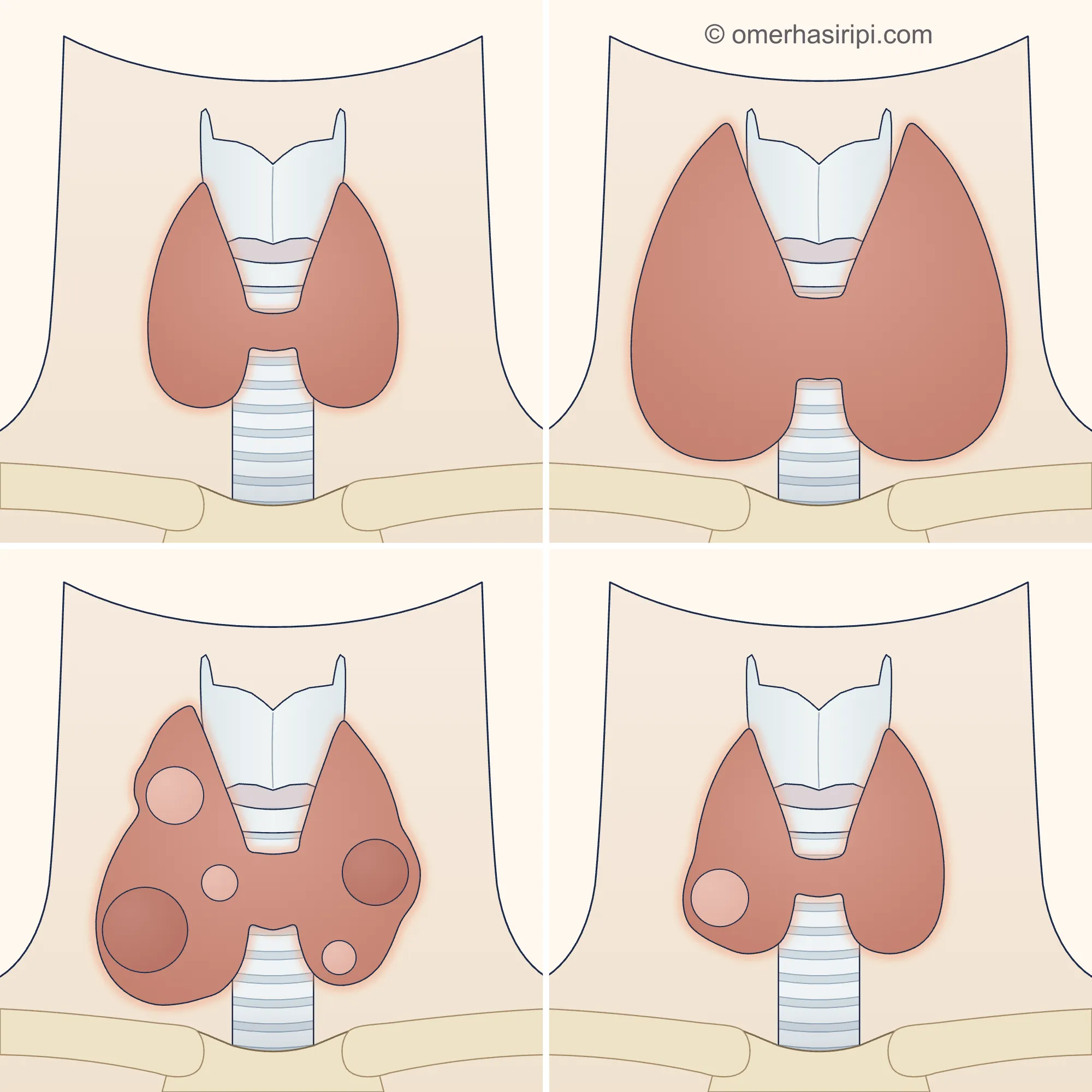
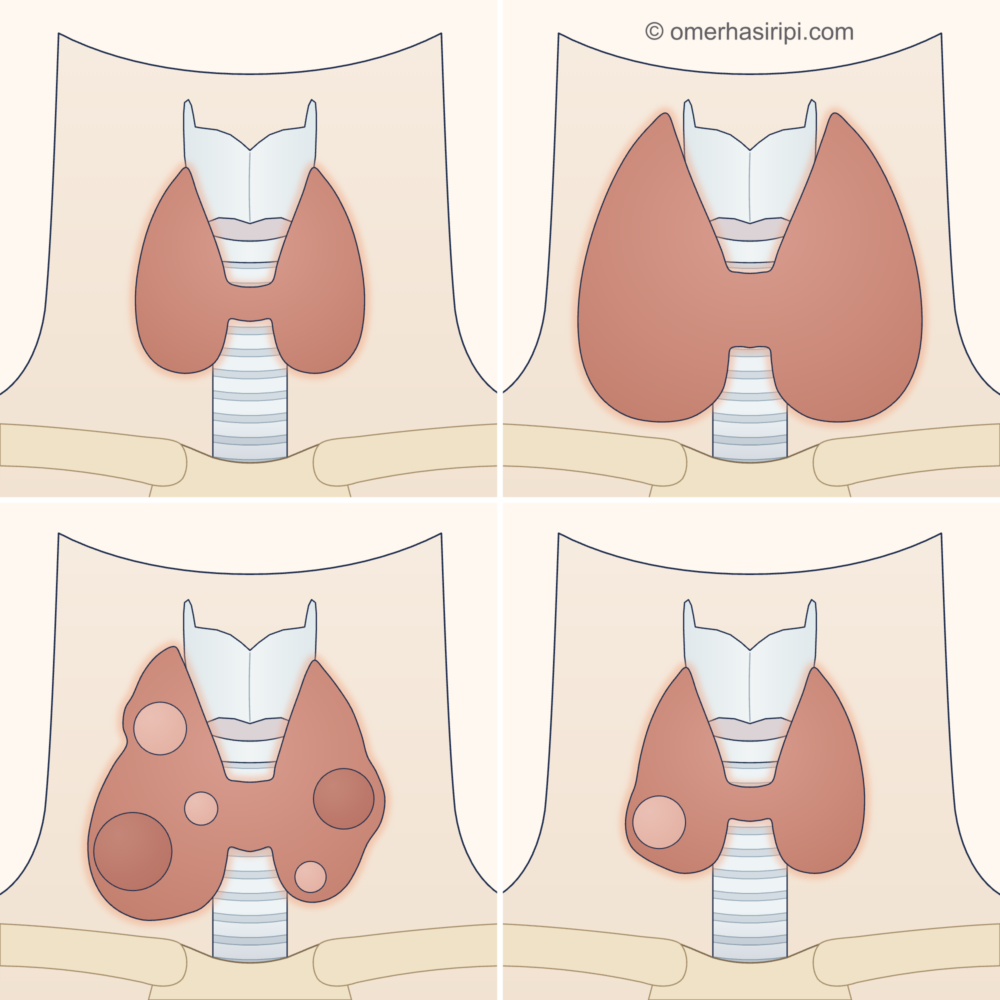
What are goitre and thyroid nodules?
Goitre is when the thyroid gland is larger than it should be for a person's age and sex[1]. The gland may enlarge as a whole (diffuse goitre); one or more nodules may also form in the gland over the years (nodular goitre; multinodular goitre if there is more than one nodule)[1]. A thyroid nodule is a lump within the gland that appears distinct from the surrounding tissue on ultrasound[1, 2].
Nodules are very common: an ultrasound assessment in Turkey reported nodules in 23.5% of people aged 18–65 and 37% of those over 65[1].
Most nodules are benign[2]. The cancer rate varies according to the group studied: according to the Turkish guideline, 5% of nodules found by ultrasound in the general population are cancerous; according to the American guideline, 7–15% of nodules assessed by a doctor are cancerous[1, 3]. When cancer is found, most are small, slow-growing types[2]. Having more than one nodule does not increase the likelihood of cancer compared with having a single nodule[1, 3].
What causes goitre and thyroid nodules?
What are the symptoms of goitre and thyroid nodules?
Most benign nodules and simple goitres cause no symptoms; they are often noticed by chance[1, 2]. As a goitre grows, it may be seen as a swelling at the front of the neck that moves when you swallow; this swelling is often painless[1, 11].
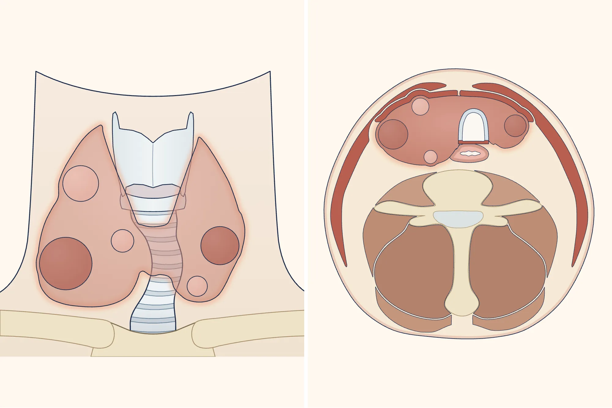
A large goitre can press on the oesophagus, windpipe and nerves that enable the voice, causing difficulty swallowing, shortness of breath and hoarseness[1]. A persistent cough, a feeling that something is stuck in the throat and wheezing when breathing in may also occur[11].
Enlarge
Swipe the drawing right or left to see the details. You can use the arrow keys on your keyboard.
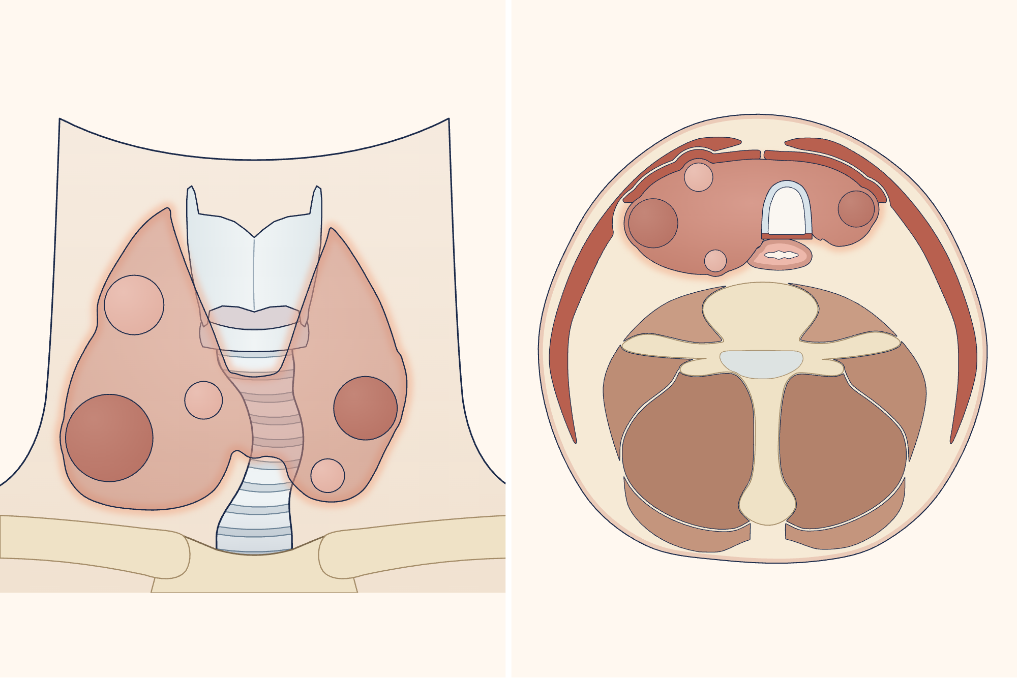
- 1 Enlarged thyroid gland (goitre)
- 2 Windpipe narrowed by pressure
- 3 Oesophagus under pressure
- 4 Voice box
- 5 Spine
Description of the drawing
An enlarged thyroid gland (goitre) grows in front of and on both sides of the windpipe. A large goitre can squeeze the windpipe from the sides and push it sideways, and can also press on the oesophagus behind it; this can make breathing and swallowing difficult. The drawing on the right is a cross-section of the neck; the front of the neck is at the top.
Illustrative drawing, not to scale. Source: Tikaa / Wikimedia Commons, CC BY-SA 4.0. The cross-section in the right panel has been adapted: letters, coloured fascia lines and two bundles of blood vessels and nerves were removed; the drawing was recoloured; the thyroid gland was enlarged and nodules added, the neck muscles and spinal canal were redrawn, the windpipe was compressed and pushed sideways, and the oesophagus was flattened. The front view in the left panel is an original drawing created for this site. Numbered labels were added. This adaptation is also licensed under CC BY-SA 4.0.

Over time, nodules may start to produce excess hormone (toxic nodular goitre); a fast heartbeat, sweating, weight loss, intolerance to heat and irritability may occur in this case[1].
Can a thyroid nodule cause pain?
Pain is rare[1, 2]. A painful swelling that appears suddenly in the neck is often due to bleeding into the nodule; inflammation of the thyroid (thyroiditis) can cause a similar picture[1]. A painful swelling that grows progressively larger may be serious[1].
Which department treats goitre and thyroid nodules?
The endocrinology department usually carries out the initial assessment; your family doctor or workplace doctor can also perform the first examination and, if they consider it necessary, refer you to endocrinology. If surgery is needed, the general surgery department performs it.
Why is a thyroid biopsy performed, and who needs one?
The first blood test is TSH, which shows how the thyroid is working; the first imaging method is an ultrasound of the neck[1]. If TSH is low, scintigraphy (a thyroid scan) is used to see whether the nodule is a “hot” nodule that produces too much hormone[1, 2]; a biopsy is not needed for a hot nodule because the likelihood of cancer is considered very low[1].
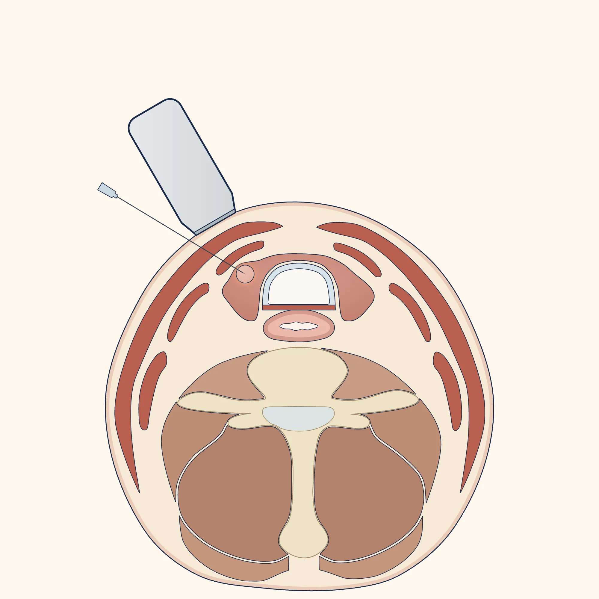
Not every nodule needs a biopsy[3]. The decision to perform a fine needle aspiration biopsy (taking a sample of cells from the nodule with a fine needle) depends on both the nodule’s appearance on ultrasound and its size[1, 2]. According to the European system (EU-TIRADS) recommended by the Turkish guideline, a biopsy is performed on the following nodules (and on smaller ones if there is a suspicious lymph node or suspected extension beyond the gland)[1, 2]:
- EU-TIRADS 3 (2–4% likelihood of cancer): if larger than 20 mm,
- EU-TIRADS 4 (6–17%): if larger than 15 mm,
- EU-TIRADS 5 (26–87%): if larger than 10 mm[1, 2]; also at 5–10 mm if close to the windpipe or the nerve that controls the voice[2].
For EU-TIRADS 2 nodules that appear benign (for example, a cyst containing only fluid), a biopsy is not needed unless treatment is planned[2, 3]. The American guideline uses a different system and different size thresholds[3].
Enlarge
Swipe the drawing right or left to see the details. You can use the arrow keys on your keyboard.
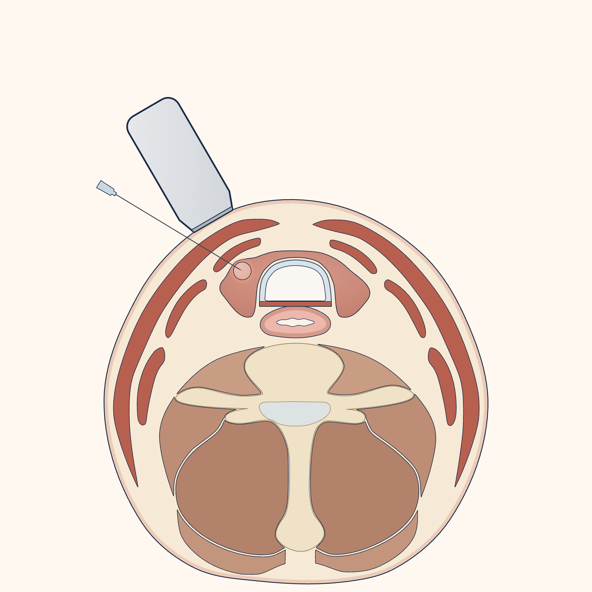
- 1 Fine needle
- 2 Nodule
- 3 Thyroid gland
- 4 Ultrasound probe
- 5 Windpipe
- 6 Oesophagus
Description of the drawing
In a fine needle aspiration biopsy, an ultrasound probe is placed on the skin of the neck and shows the nodule; a fine needle is advanced through the skin into the nodule and a sample of cells is taken from it. The drawing is a cross-section of the neck; the front of the neck is at the top.
Results may vary from person to person with any surgical or interventional procedure. You are advised to discuss the procedure in detail with your doctor beforehand.
Illustrative drawing, not to scale. Source: Tikaa / Wikimedia Commons, CC BY-SA 4.0. Letters, coloured fascia lines and two bundles of blood vessels and nerves were removed; the drawing was recoloured; the windpipe, oesophagus, spinal canal and thyroid gland were redrawn; a thyroid nodule, ultrasound probe and fine needle were added; numbered labels were added. This adaptation is also licensed under CC BY-SA 4.0.
How is a thyroid biopsy performed?
The biopsy is performed under ultrasound guidance[1, 2]. According to the Turkish guideline, local anaesthesia (numbing the area) is not necessary for the procedure[1]. During or after the procedure, although very rare, significant bleeding that narrows the airway may occur; this possibility should be explained to you before the procedure[1, 5]. If your neck swells rapidly after the procedure, go to the emergency department without delay; if you have difficulty breathing, call 112[5, 11]. Taking blood-thinning medication does not in itself prevent a biopsy[2, 5]. If you take these medicines, tell your doctor; do not stop taking them on your own[5].
What does the biopsy result mean?
The result is reported in one of six groups (the Bethesda system)[1]. The percentage in brackets is the average likelihood of cancer for that group according to the Turkish guideline[1]:
- Inadequate sample (13%): the biopsy is repeated.
- Benign (4%): monitored with ultrasound.
- Atypia of uncertain significance (22%): repeat biopsy, molecular (genetic) testing, removal of half the thyroid (lobectomy) or monitoring.
- Follicular neoplasm (30%): molecular testing or lobectomy.
- Suspicious for cancer (74%): molecular testing, lobectomy or removal of the whole thyroid.
- Cancer (97%): lobectomy or removal of the whole thyroid[1].
What should people with thyroid nodules be aware of?
For nodules that cause no symptoms, the interval between checks depends on the nodule’s ultrasound appearance and biopsy result; guidelines recommend different intervals[1, 2, 3]. According to the European guideline, for nodules that cause no symptoms and do not need a biopsy: no further check is needed for a 5–10 mm EU-TIRADS 2–3 nodule; EU-TIRADS 2 nodules larger than 10 mm and EU-TIRADS 3 nodules of 10–20 mm are checked after 3–5 years, EU-TIRADS 4 nodules smaller than 15 mm after 1 year, and EU-TIRADS 5 nodules smaller than 10 mm every 6–12 months; if an EU-TIRADS 5 nodule remains unchanged for two years, checks can be less frequent[2]. These intervals do not apply to nodules where uptake in one area of the thyroid is found incidentally on an FDG PET-CT scan performed for another reason; according to the Turkish guideline, these nodules are assessed with ultrasound, a biopsy is performed if they are EU-TIRADS 4–5 and larger than 10 mm or EU-TIRADS 3 and larger than 20 mm, and EU-TIRADS 2–3 nodules smaller than 20 mm are monitored with ultrasound every 6–12 months; your doctor decides how the others are monitored[1]. For nodules with one benign biopsy result that do not look highly suspicious on ultrasound, the interval between checks ranges from 12–24 months to 3–5 years; for those that look highly suspicious, the biopsy is repeated within one year[1, 2, 3]. According to the American guideline, a nodule whose second biopsy is also benign does not need ultrasound monitoring for cancer[3]. Nodules that cause symptoms but are not treated are reassessed within one year[2]. For simple goitre, checks are initially carried out once a year[1].
Even if a benign nodule grows a little, it is usually only monitored if it causes no symptoms; if it grows significantly, a repeat biopsy is considered[2, 3]. If your symptoms change, get worse or new symptoms start, see your doctor without waiting for your next check[2, 4, 12].
Tell your doctor if you received radiotherapy to your head or neck as a child, or if your mother, father, sibling or child has thyroid cancer: these increase the likelihood of cancer[1, 2, 3].
Can goitre go away with medication, and what helps?
In people with normal thyroid hormone levels, simple goitre often does not need treatment and is only monitored[1]. Thyroid hormone medication (levothyroxine) is not recommended to shrink nodules in these people[1, 2]: its shrinking effect is small (an average of 5–15% of nodule volume), while the high doses needed increase the risk of heart rhythm problems and osteoporosis[3]. If you take this medicine, consult the doctor who prescribed it before stopping.
Iodine supplements are only needed by people with iodine deficiency[2]. For pregnant women, the Turkish guideline recommends iodine supplementation starting when pregnancy is planned and continuing until breastfeeding ends; your doctor determines the dose[1]. The same guideline recommends refined iodised table salt for everyone; it does not recommend unrefined salts of unknown composition, such as rock salt and gourmet salts[1]. Iodised salt is of little benefit to a person who has had a goitre for a long time, but it benefits other family members, especially younger ones[1]. The guidelines do not recommend any food, plant or natural method to shrink a goitre or nodule[1].
When is surgery needed for a goitre or thyroid nodule?
If a goitre presses on the windpipe or oesophagus and causes symptoms, surgery is recommended[1, 7]; if a nodule causes pressure symptoms, surgery is one of the options[2].
Surgery is also considered in the following situations:
- if the biopsy result is cancer or suspicious for cancer, or if the result is uncertain and unsuitable for monitoring[2],
- if the nodule is 4 cm or larger, even if the biopsy is benign, because the likelihood of a biopsy being wrong increases at this size (European guideline)[2],
- if the goitre extends behind the collarbone or breastbone[2],
- if its appearance bothers you[1, 2],
- if the nodule or goitre produces too much hormone[2].
Studies have shown that breathing and swallowing improve after surgery for a goitre causing pressure symptoms[7].
How is goitre surgery performed, and how long does it take?
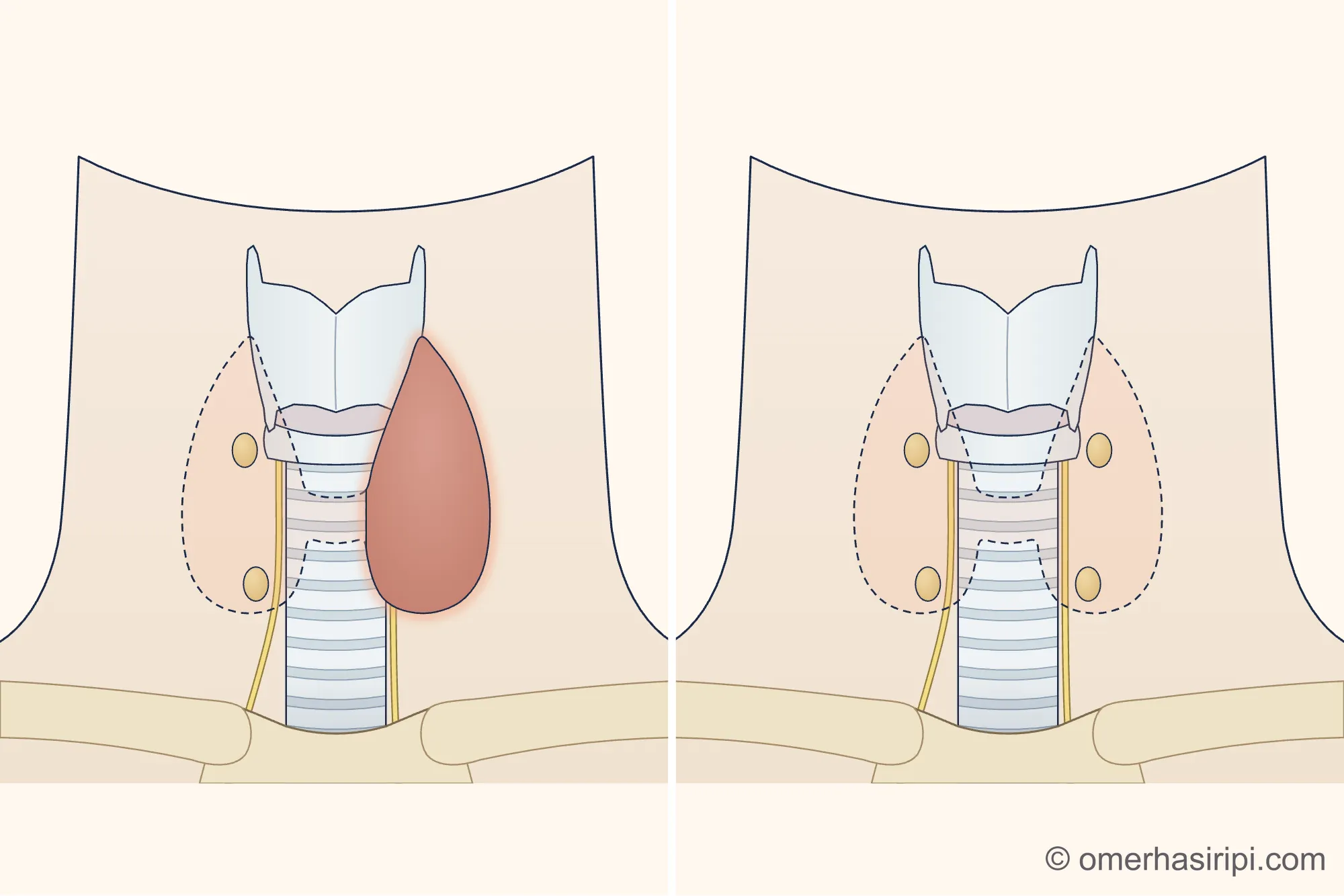
Depending on the condition, half the thyroid (lobectomy) or the whole thyroid is removed[1, 2]. The operation is performed under a general anaesthetic; an incision is made along a skin crease in the lower neck[17]. During the operation, care is taken to preserve the small glands behind the thyroid that regulate calcium (parathyroid glands) and the nerve that controls the voice[17]. The operation usually takes 2–4 hours; the time varies with the size of the thyroid[17]. Most people return to work after 1–2 weeks; this may take longer for heavy physical work[14, 17]. Before driving, you should be able to turn your neck comfortably and brake suddenly; most surgeons recommend waiting at least 3–5 days[14, 17].
Enlarge
Swipe the drawing right or left to see the details. You can use the arrow keys on your keyboard.
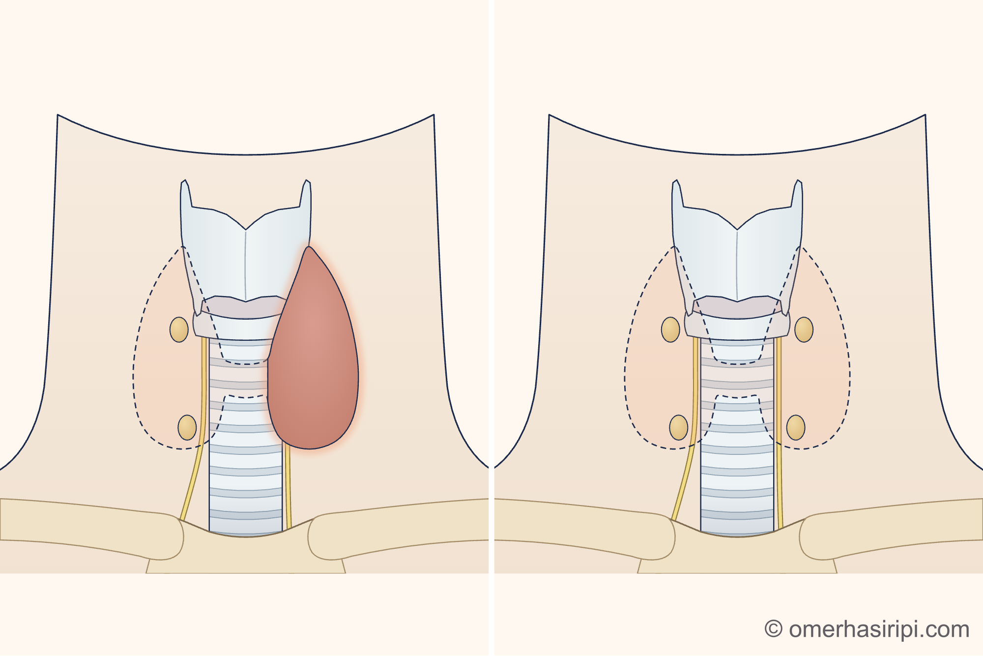
- 1 Half of the thyroid removed (lobectomy)
- 2 Remaining half
- 3 Whole thyroid removed
- 4 Small glands that regulate calcium (parathyroid glands)
- 5 Nerve that controls the voice
- 6 Windpipe
- 7 Voice box
Description of the drawing
Depending on the condition, half the thyroid (lobectomy; left) or the whole thyroid (right) is removed; the part removed is shown by a dashed line. The small glands that regulate calcium (parathyroid glands; usually two on each side) lie just behind the thyroid, and the nerve that controls the voice lies on each side of the windpipe. Care is taken to preserve these during surgery; because they are very close to the gland, they can be affected by the operation. The path of the nerve is shown approximately in the drawing.
Results may vary from person to person with any surgical or interventional procedure. You are advised to discuss the procedure in detail with your doctor beforehand.
Illustrative drawing, not to scale. Prepared for this site.
What treatments are available besides surgery?
Radioactive iodine is recommended as an alternative to surgery for nodules that produce too much hormone; it may also be considered for people who cannot or do not want to have surgery for a benign goitre with several nodules (multinodular goitre) that causes symptoms despite normal hormone levels[1, 2]. It is not often used for simple goitre in Turkey[1]. Radioactive iodine treatment is not given during pregnancy; women of childbearing age have a pregnancy test before treatment[1, 6]. Breastfeeding and expressing milk must stop before treatment (at least 6 weeks according to the Turkish guideline, at least 3 months according to the American guidelines)[1, 6, 16] and must not be restarted for this baby[15]; after treatment, women should not become pregnant for at least 6 months and until their thyroid function has stabilised[1, 6], and men should postpone having a child for 3–4 months[1]. Avoid close contact with young children and pregnant women for a time after treatment; your team will tell you for how long[15]. Hormone deficiency can develop over time after radioactive iodine; lifelong checks are recommended[1, 2]. During pregnancy, scintigraphy is usually postponed until after childbirth; if you are breastfeeding, say so before the procedure: depending on the substance used, you may need to pause breastfeeding for a time or stop breastfeeding[1].
In ablation, alcohol is injected under ultrasound guidance into benign cysts that do not resolve with drainage; benign solid nodules that cause symptoms are shrunk with heat[2, 5]. The Turkish ablation guideline considers this a first-line alternative to surgery for a single or dominant benign nodule that causes pressure symptoms or concerns about appearance[5]. After ablation, if you develop severe or increasing pain, rapidly growing swelling in the neck, redness, fever, a change in your voice or difficulty swallowing, contact the team that performed the procedure or go to the emergency department without delay; if you have difficulty breathing, call 112[5, 11].
Is goitre surgery risky, and what happens afterwards?
Before surgery, your doctor should discuss possibilities such as a change in voice, low calcium, bleeding, a scar, the condition returning and the need for hormone medication[6].
The nerve that controls the voice can be affected after surgery[1]. According to figures provided by the American ear, nose and throat association for thyroid surgery, this nerve is temporarily affected in about 1 in 10 people, and voice problems last longer in up to 1 in 25 people[10]. Most nerve problems usually improve within a few months[6]. Your voice should be assessed after surgery, usually between the second week and the second month, and if your voice is affected, your vocal cords should be examined; a problem detected early can be treated early[6].
If the small glands that regulate calcium (parathyroid glands) are affected, blood calcium levels may fall[1]; this is the most common problem after surgery on both sides of the thyroid[9]. Mild or moderate low calcium can be treated with calcium and vitamin D taken by mouth; severe cases may need treatment in hospital[9].
If the whole thyroid has been removed, lifelong thyroid hormone medication is needed[1]. Hormone deficiency can also develop when half is removed: after lobectomy for nodules that do not produce too much hormone, this has been reported in up to 20–30% of people[1]. Some people experience neck tightness that is felt most during the first 3 months; massage and neck movements may help in the early period[6].
Sources
19
- Türkiye Endokrinoloji ve Metabolizma Derneği (TEMD), Tiroid Çalışma Grubu. Tiroid Hastalıkları Tanı ve Tedavi Kılavuzu 2025. 8th edition. Ankara: TEMD; 2025. Text: file.temd.org.tr.
- Durante C, Hegedüs L, Czarniecka A, Paschke R, Russ G, Schmitt F, et al. 2023 European Thyroid Association Clinical Practice Guidelines for thyroid nodule management. Eur Thyroid J. 2023;12(5):e230067. doi: 10.1530/ETJ-23-0067. PubMed: 37358008.
- Haugen BR, Alexander EK, Bible KC, Doherty GM, Mandel SJ, Nikiforov YE, et al. 2015 American Thyroid Association Management Guidelines for Adult Patients with Thyroid Nodules and Differentiated Thyroid Cancer. Thyroid. 2016;26(1):1–133. doi: 10.1089/thy.2015.0020. PubMed: 26462967.
- National Institute for Health and Care Excellence (NICE). Thyroid disease: assessment and management. NICE guideline NG145. 2019; last updated 12 October 2023. Text: www.nice.org.uk.
- Türkiye Endokrinoloji ve Metabolizma Derneği (TEMD). Tiroid ve Paratiroid Lezyonlarında Girişimsel İşlemler ve Ablasyon Tedavileri Kılavuzu 2026. Ankara: TEMD; 2026. Text: file.temd.org.tr.
- Ringel MD, Sosa JA, Baloch Z, Bischoff L, Bloom G, Brent GA, et al. 2025 American Thyroid Association Management Guidelines for Adult Patients with Differentiated Thyroid Cancer. Thyroid. 2025;35(8):841–985. doi: 10.1177/10507256251363120. PubMed: 40844370.
- Chen AY, Bernet VJ, Carty SE, Davies TF, Ganly I, Inabnet WB, et al. American Thyroid Association statement on optimal surgical management of goiter. Thyroid. 2014;24(2):181–189. (Only the abstract could be read.) doi: 10.1089/thy.2013.0291. PubMed: 24295043.
- Terris DJ, Snyder S, Carneiro-Pla D, Inabnet WB, Kandil E, Orloff L, et al. American Thyroid Association statement on outpatient thyroidectomy. Thyroid. 2013;23(10):1193–1202. (Only the abstract could be read.) doi: 10.1089/thy.2013.0049. PubMed: 23742254.
- Orloff LA, Wiseman SM, Bernet VJ, Fahey TJ, Shaha AR, Shindo ML, et al. American Thyroid Association Statement on Postoperative Hypoparathyroidism: Diagnosis, Prevention, and Management in Adults. Thyroid. 2018;28(7):830–841. (Only the abstract could be read.) doi: 10.1089/thy.2017.0309. PubMed: 29848235.
- Chandrasekhar SS, Randolph GW, Seidman MD, Rosenfeld RM, Angelos P, et al. Clinical practice guideline: improving voice outcomes after thyroid surgery. Otolaryngol Head Neck Surg. 2013;148(6 Suppl):S1–37. (Only the abstract could be read.) doi: 10.1177/0194599813487301. PubMed: 23733893.
- NHS (United Kingdom National Health Service). Goitre (goitre): patient information page. Last reviewed: 11 March 2026. Text: www.nhs.uk.
- NHS (United Kingdom National Health Service). Symptoms of thyroid cancer (symptoms of thyroid cancer): patient information page. Last reviewed: 18 May 2023. Text: www.nhs.uk.
- Iliff HA, El-Boghdadly K, Ahmad I, Davis J, Harris A, Khan S, et al. Management of haematoma after thyroid surgery: systematic review and multidisciplinary consensus guidelines from the Difficult Airway Society, the British Association of Endocrine and Thyroid Surgeons and the British Association of Otorhinolaryngology, Head and Neck Surgery. Anaesthesia. 2022;77(1):82–95. doi: 10.1111/anae.15585. PubMed: 34545943.
- American Thyroid Association (ATA). Post Operative Expectations: patient information page. No date on the page (© 2026). Accessed: 28 September 2026. Text: www.thyroid.org.
- American Thyroid Association (ATA). Radioactive Iodine: patient information page. No date on the page (© 2026). Accessed: 29 September 2026. Text: www.thyroid.org.
- Dorris K. American Thyroid Association Task Force Updates Treatment Guidelines for Hyperthyroidism (patient summary of the American Thyroid Association’s 2016 hyperthyroidism guideline). Clinical Thyroidology for the Public. 2016;9(11):3–7. Accessed: 29 September 2026. Text: www.thyroid.org.
- NHS Tayside (Scotland), Ear, Nose and Throat Department. Thyroidectomy: Outpatient information (thyroid surgery: patient leaflet, LN0942). Reviewed: July 2026. Accessed: 29 September 2026. Text: www.nhstaysidecdn.scot.nhs.uk.
- Turner J, Gittoes N, Selby P; Society for Endocrinology Clinical Committee. Society for Endocrinology Endocrine Emergency Guidance: Emergency management of acute hypocalcaemia in adult patients. Endocrine Connections. 2016;5(5):G7–G8. Accessed: 6 October 2026. Text: doi.org.
- NHS (United Kingdom National Health Service). Hypoparathyroidism (underactive parathyroid glands): patient information page. Last reviewed: 10 April 2025. Text: www.nhs.uk.
Medical review of the Turkish original: Op. Dr. Ömer Seyfullah Hasıripi, . This page is a translation checked by AI reviewers; the Turkish version prevails.
This page is for general information; it does not replace an examination. See a doctor about your symptoms.