Is surgery necessary, how is it done and how long is recovery?

Groin hernia (inguinal hernia)

In short

A groin hernia is when fatty tissue or bowel from inside the abdomen pushes out through a weak spot in the groin. It does not go away on its own. It can only be repaired with surgery. However, watching and waiting is also an option for men with few or no symptoms. It is an emergency if the hernia suddenly becomes very painful or no longer goes back in when it previously did.

Conditions covered on this page

Treatment options

On this page
Forms of a groin hernia

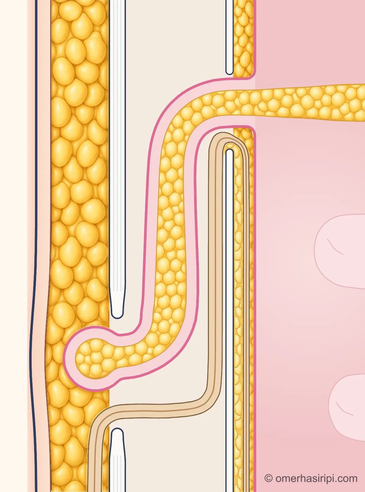
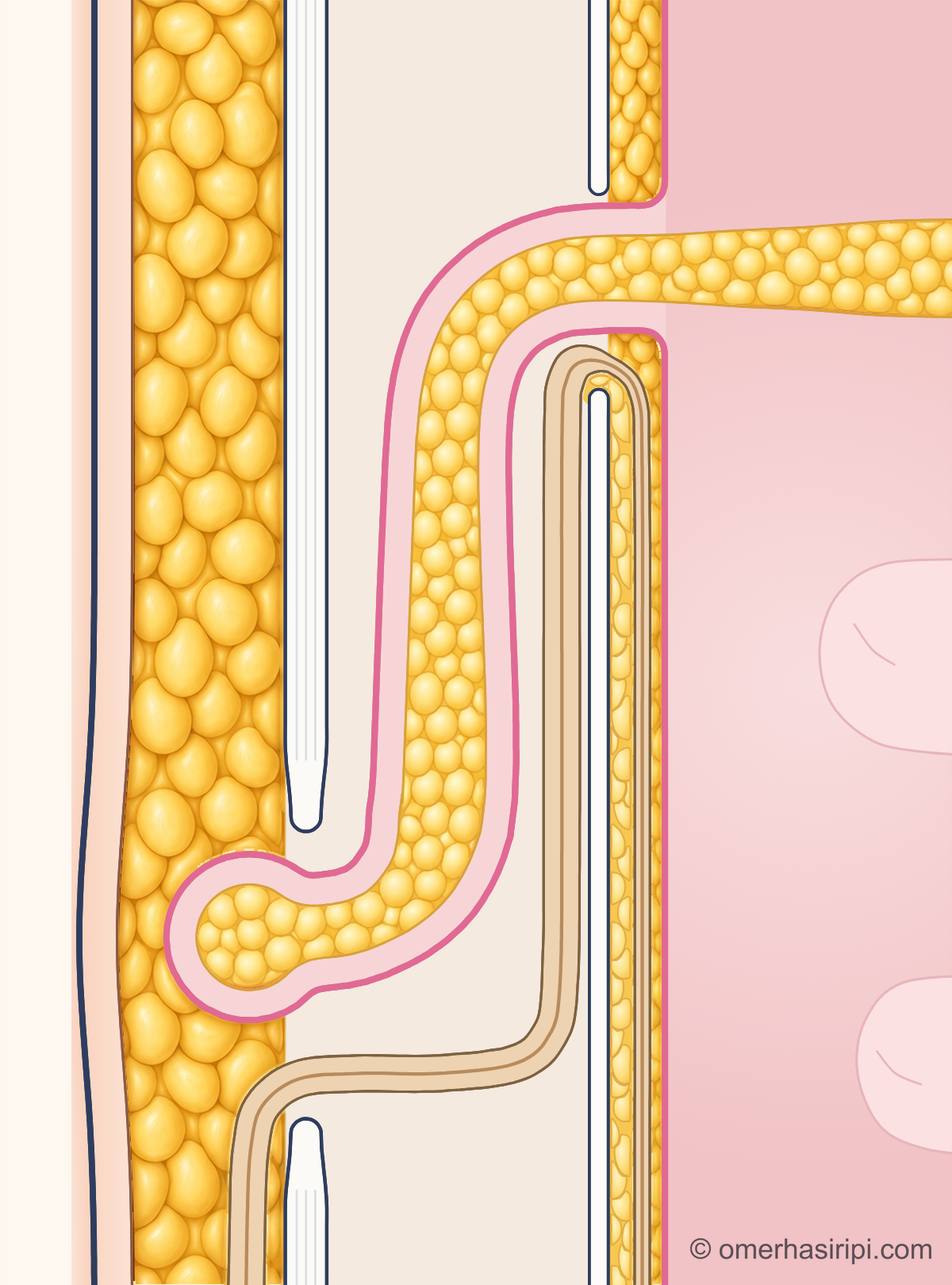
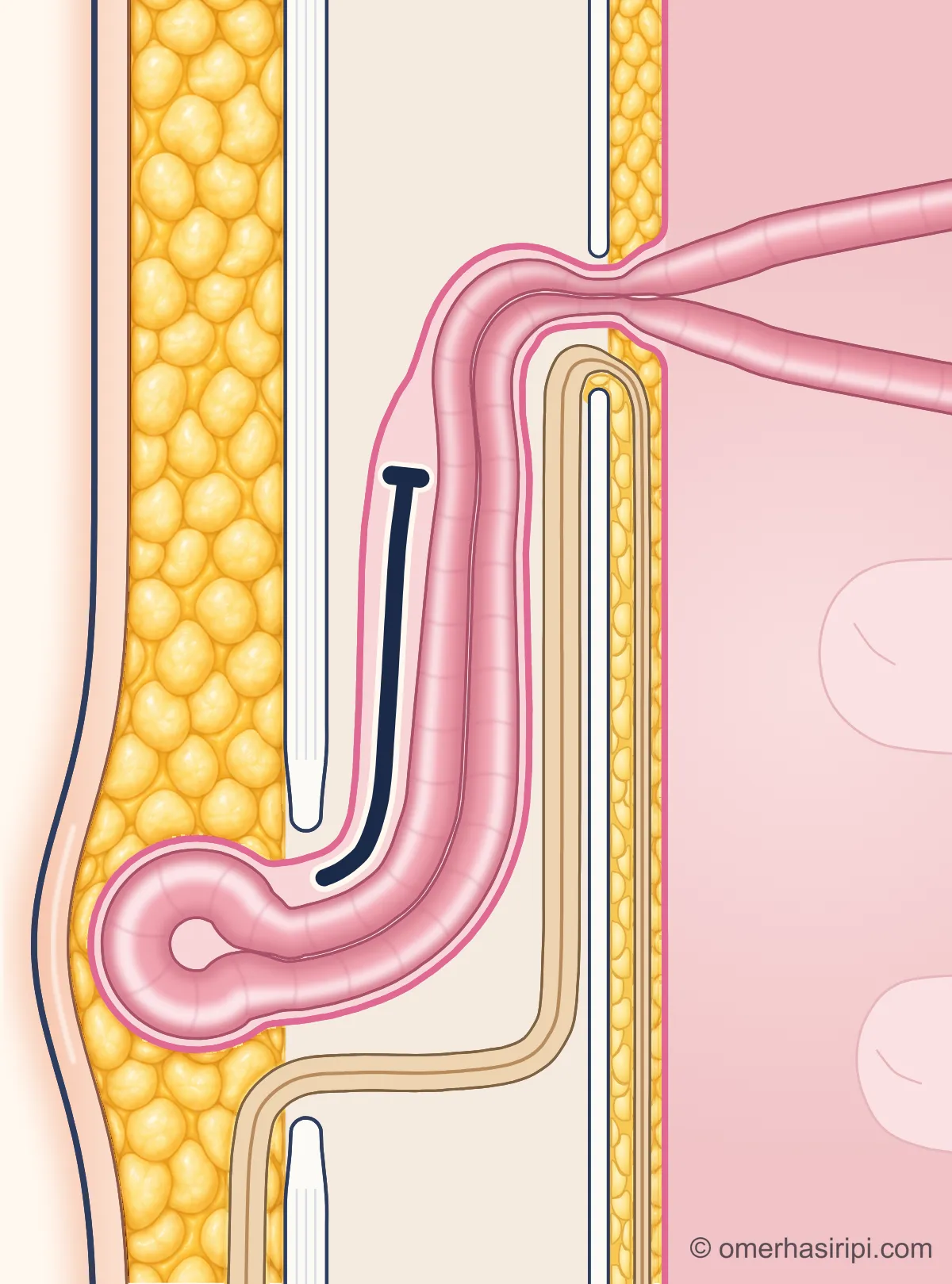
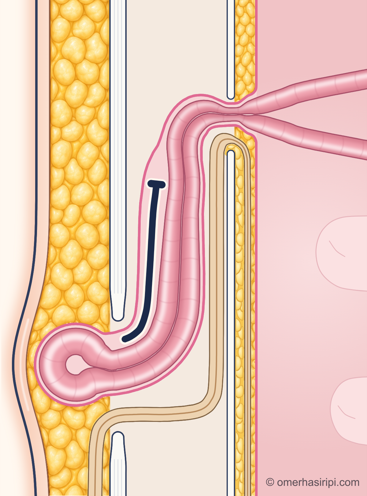
The sections look along the right inguinal canal: the skin is on the left and the inside of the abdomen on the right; the upper end is towards the hip and the lower end towards the pubic bone. The type drawn is an indirect inguinal hernia: it enters through the inner opening and runs down along the canal; a direct inguinal hernia pushes forward through a weak spot in the back wall of the canal. Fatty tissue is not a stage: a hernia may also contain fat instead of bowel.

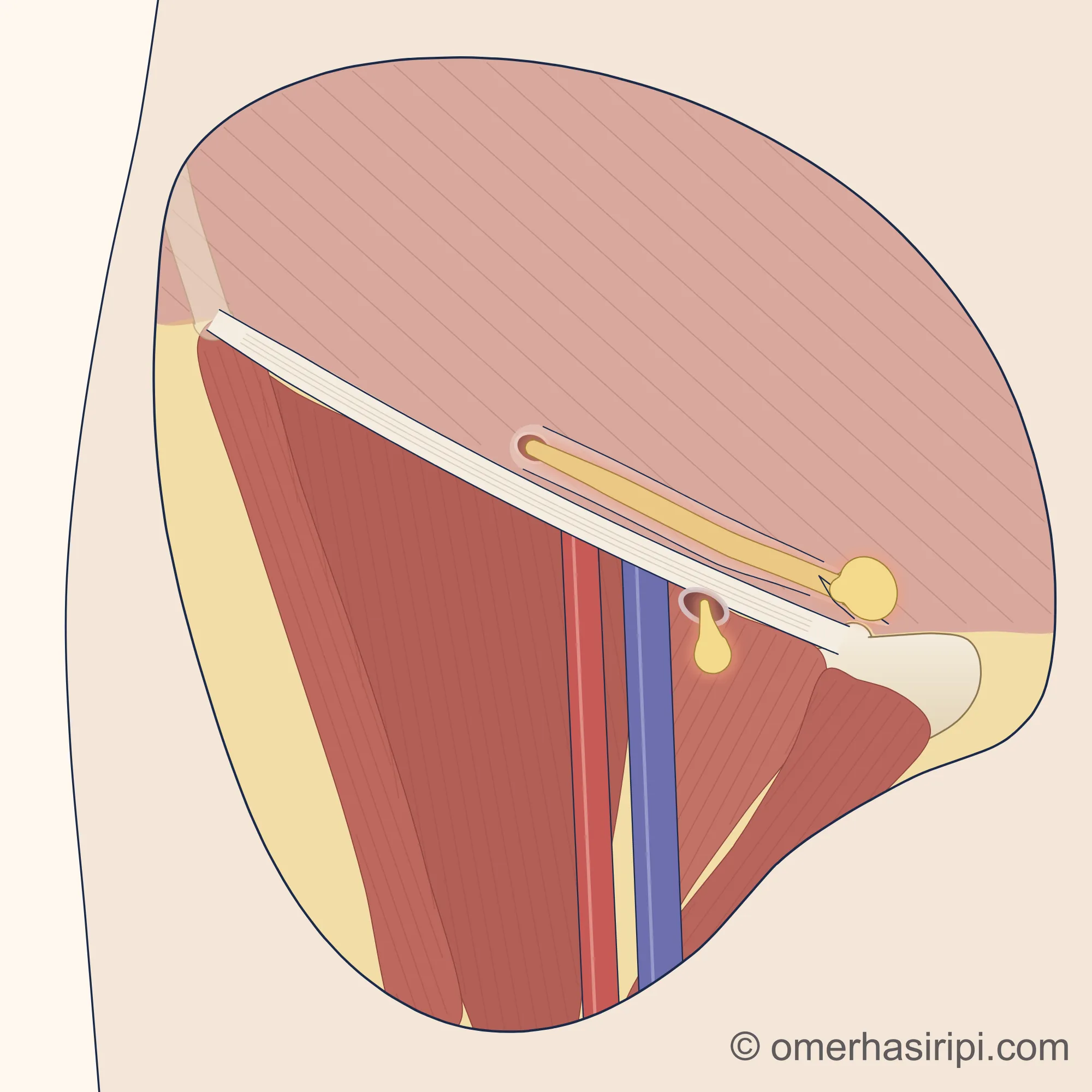
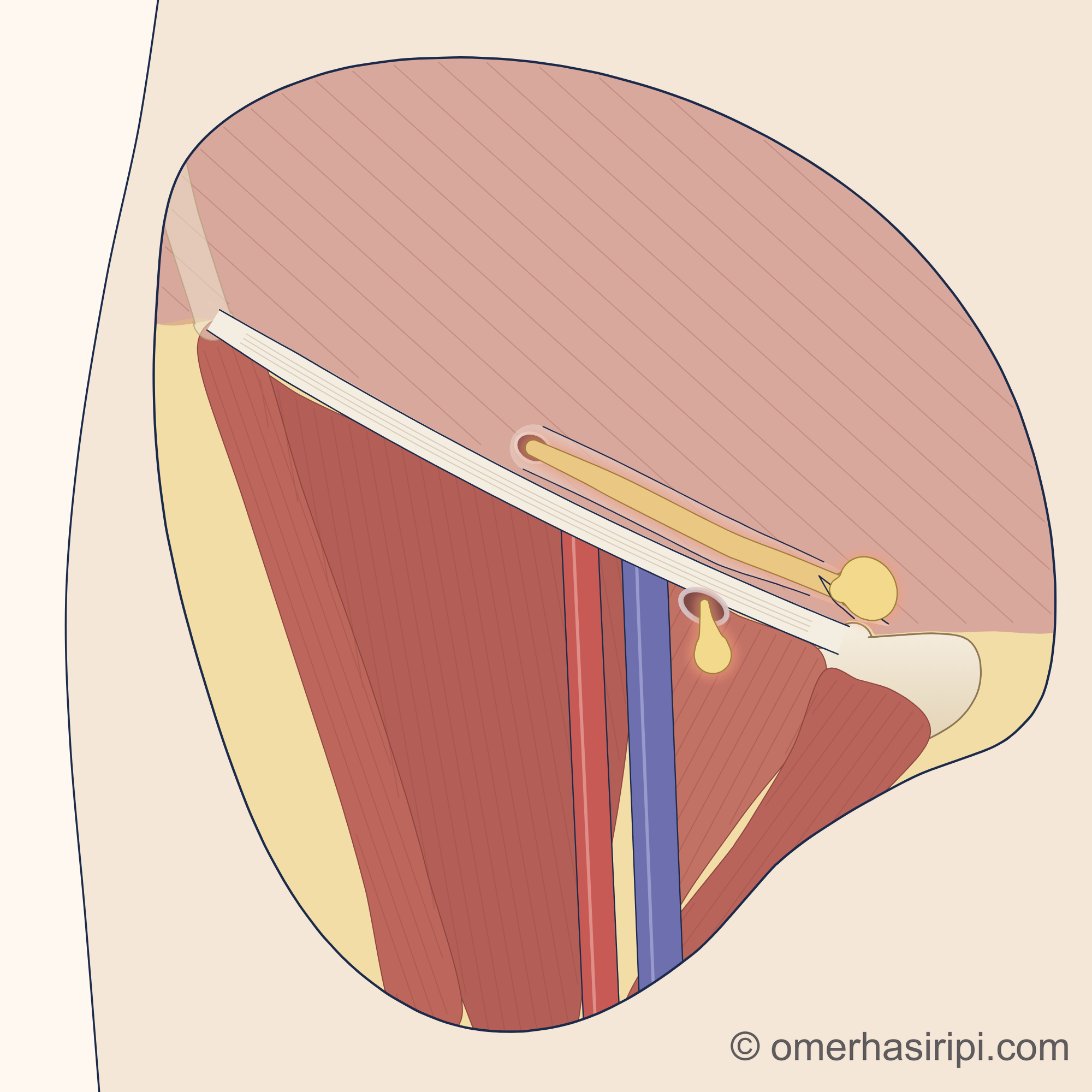
A close-up schematic drawing of the right groin viewed from the front: the hip is on the left and the midline on the right. The skin and underlying fatty tissue have been removed within a round window; the area outside the window is flat and pale. A flat, pale, strong band (the inguinal ligament) runs downwards and inwards from the bony prominence at the front of the hip bone at the upper left to the prominence of the pubic bone beside the midline on the right; the band ends at this prominence, and the pubic bone is visible from the prominence towards the midline as a separate, pale ivory-coloured bone. Just above the inner half of the ligament, the inguinal canal is shown as a transparent tube running obliquely and parallel to it: a soft sac of fat (groin hernia) enters through its inner opening at the level of the middle of the ligament, travels along the canal in one continuous piece and bulges out as a round sac through a small triangular slit in the membrane of the outermost muscle layer of the abdominal wall, just above the pubic bone prominence. The red main artery runs down to the leg beneath the ligament on the outer side, with the blue main vein on its inner side; the inner opening of the canal lies above and to the outer side of the artery. On the inner side of the vein, a smaller, round sac of fat (femoral hernia) hangs down through a small opening surrounded by a pale border just below the ligament (the opening of the femoral canal).
Enlarge

Swipe the drawing right or left to see the details. You can use the arrow keys on your keyboard.

A close-up schematic drawing of the right groin viewed from the front: the hip is on the left and the midline on the right. The skin and underlying fatty tissue have been removed within a round window; the area outside the window is flat and pale. A flat, pale, strong band (the inguinal ligament) runs downwards and inwards from the bony prominence at the front of the hip bone at the upper left to the prominence of the pubic bone beside the midline on the right; the band ends at this prominence, and the pubic bone is visible from the prominence towards the midline as a separate, pale ivory-coloured bone. Just above the inner half of the ligament, the inguinal canal is shown as a transparent tube running obliquely and parallel to it: a soft sac of fat (groin hernia) enters through its inner opening at the level of the middle of the ligament, travels along the canal in one continuous piece and bulges out as a round sac through a small triangular slit in the membrane of the outermost muscle layer of the abdominal wall, just above the pubic bone prominence. The red main artery runs down to the leg beneath the ligament on the outer side, with the blue main vein on its inner side; the inner opening of the canal lies above and to the outer side of the artery. On the inner side of the vein, a smaller, round sac of fat (femoral hernia) hangs down through a small opening surrounded by a pale border just below the ligament (the opening of the femoral canal).
  1. 1 Inguinal canal (canalis inguinalis)
  2. 2 Groin hernia (hernia inguinalis)
  3. 3 Opening of the femoral canal (anulus femoralis)
  4. 4 Femoral hernia (hernia femoralis)
  5. 5 Inguinal ligament (ligamentum inguinale)
  6. 6 Main artery of the leg (arteria femoralis)
  7. 7 Main vein of the leg (vena femoralis)
  8. 8 Pubic bone (os pubis)
Description of the drawing

Right groin, viewed from the front: the hip is on the left and the midline on the right. A groin hernia emerges above the inguinal ligament; the drawing shows the type of hernia that travels along the inguinal canal. A femoral hernia emerges below the ligament through the femoral canal on the inner side of the main vein of the leg; the two types can be difficult to distinguish by examination, especially in women. To show the structures, the skin and underlying fatty tissue have been removed, and the abdominal wall is drawn transparent so that the inguinal canal can be seen.[1, 3, 8, 9]

Illustrative drawing, not to scale. Prepared for this site.

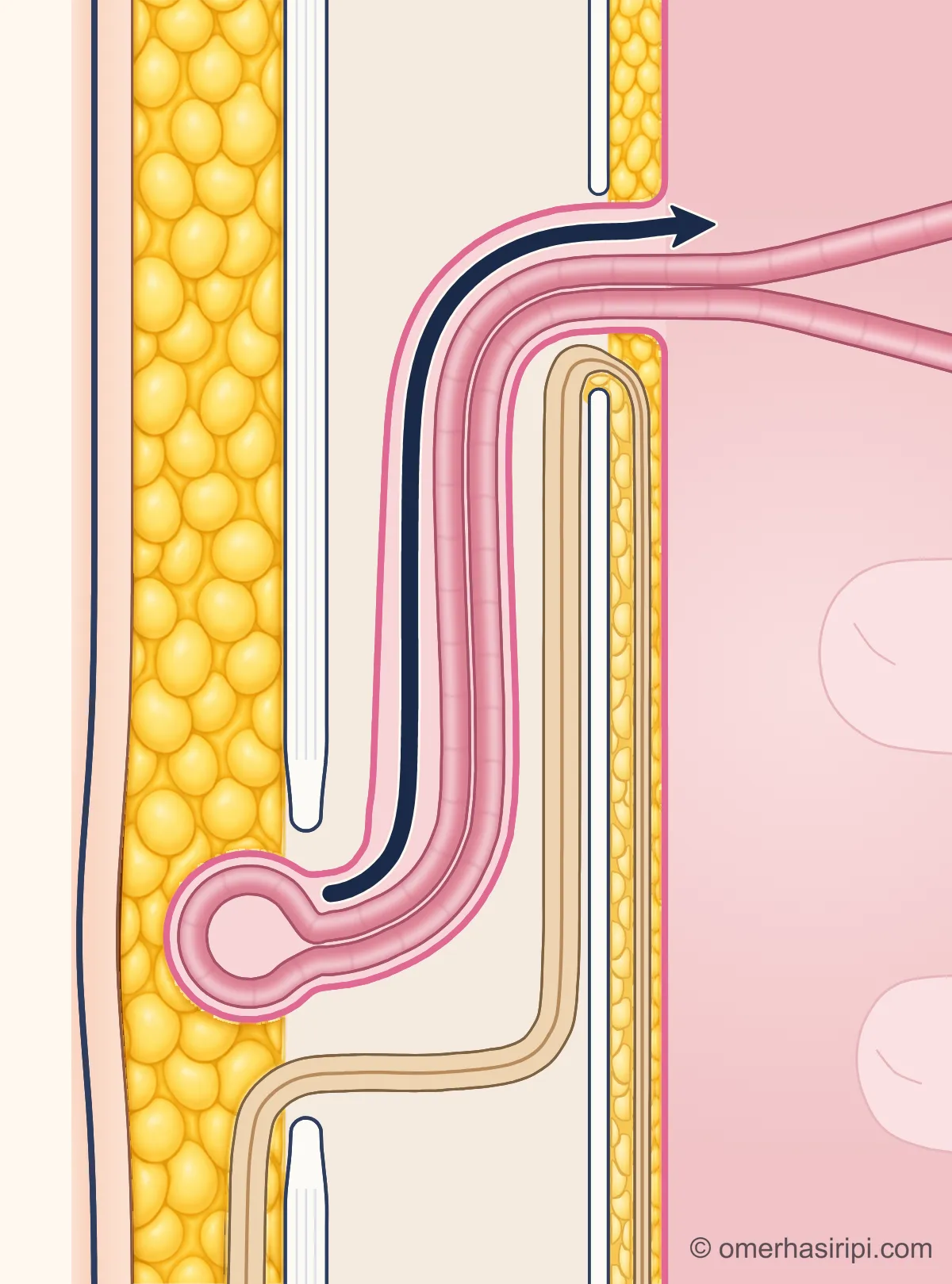
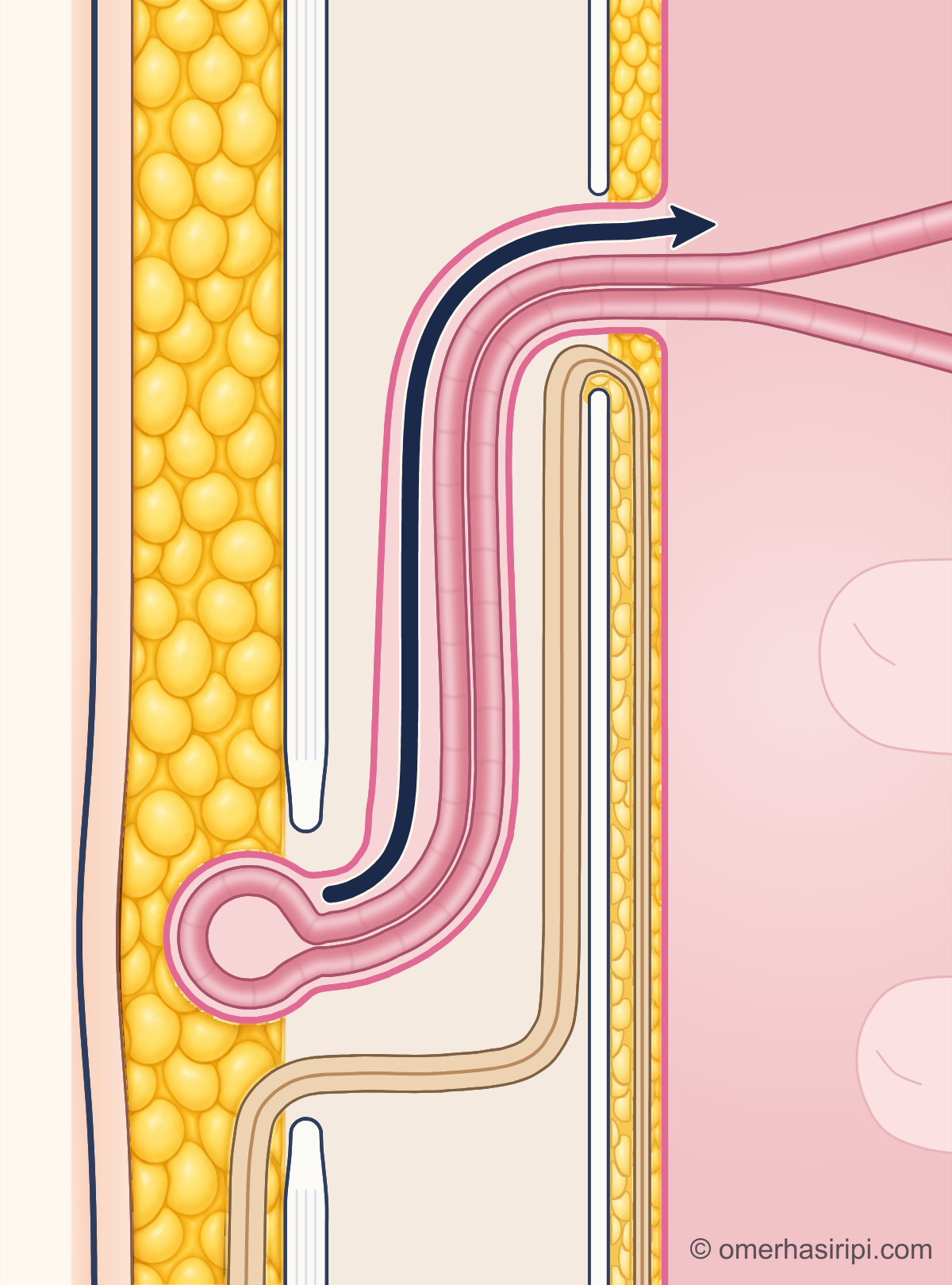
Schematic drawing: a section along the right inguinal canal of a man; the skin is on the left and the inside of the abdomen on the right, with the upper end towards the hip and the lower end towards the pubic bone. From the outside in, the drawing shows the skin, the fatty tissue beneath the skin, the tough white layer that forms the front wall of the canal, the canal itself, the thin back wall of the canal, a thin layer of fat and the abdominal lining as a pink line. The opening at the upper end of the back wall is the inner opening of the canal, and the opening at the lower end of the front wall is its outer opening. The spermatic cord enters the canal through the inner opening, runs down along the canal and comes out beneath the skin through the outer opening. A sac formed from the abdominal lining has entered the canal through the inner opening, run down on the skin side of the cord, come out beneath the skin through the outer opening and made the skin bulge. Inside the sac, a thin, pink loop of bowel lies loosely; its two limbs run up along the canal and continue into the abdomen through the inner opening. A curved navy-blue arrow runs from the sac along the canal through the inner opening towards the inside of the abdomen.
Enlarge

Swipe the drawing right or left to see the details. You can use the arrow keys on your keyboard.

Schematic drawing: a section along the right inguinal canal of a man; the skin is on the left and the inside of the abdomen on the right, with the upper end towards the hip and the lower end towards the pubic bone. From the outside in, the drawing shows the skin, the fatty tissue beneath the skin, the tough white layer that forms the front wall of the canal, the canal itself, the thin back wall of the canal, a thin layer of fat and the abdominal lining as a pink line. The opening at the upper end of the back wall is the inner opening of the canal, and the opening at the lower end of the front wall is its outer opening. The spermatic cord enters the canal through the inner opening, runs down along the canal and comes out beneath the skin through the outer opening. A sac formed from the abdominal lining has entered the canal through the inner opening, run down on the skin side of the cord, come out beneath the skin through the outer opening and made the skin bulge. Inside the sac, a thin, pink loop of bowel lies loosely; its two limbs run up along the canal and continue into the abdomen through the inner opening. A curved navy-blue arrow runs from the sac along the canal through the inner opening towards the inside of the abdomen.
  1. 1 Hernia sac (saccus herniae)
  2. 2 Small bowel pushed outwards (intestinum tenue)
  3. 3 Inner opening of the inguinal canal (anulus inguinalis profundus)
  4. 4 Can return inside
  5. 5 Outer opening of the inguinal canal (anulus inguinalis superficialis)
  6. 6 Spermatic cord (funiculus spermaticus)
Description of the drawing

A hernia may also contain bowel. A hernia that can go back in normally returns to its place easily; the swelling may get smaller when you lie down.[1, 5, 15, 16, 17]

Illustrative drawing, not to scale. Prepared for this site.

Schematic drawing: a section along the right inguinal canal of a man; the skin is on the left and the inside of the abdomen on the right, with the upper end towards the hip and the lower end towards the pubic bone. From the outside in, the drawing shows the skin, the fatty tissue beneath the skin, the tough white layer that forms the front wall of the canal, the canal itself, the thin back wall of the canal, a thin layer of fat and the abdominal lining as a pink line. The opening at the upper end of the back wall is the inner opening of the canal, and the opening at the lower end of the front wall is its outer opening. The spermatic cord enters the canal through the inner opening, runs down along the canal and comes out beneath the skin through the outer opening. A sac formed from the abdominal lining has entered the canal through the inner opening, run down on the skin side of the cord, come out beneath the skin through the outer opening and made the skin bulge. Yellow fatty tissue made up of small round lobules fills the inside of the sac, leaving a thin pink rim; this fat runs up along the canal and continues into the abdomen through the inner opening as a band. There is no bowel in the sac.
Enlarge

Swipe the drawing right or left to see the details. You can use the arrow keys on your keyboard.

Schematic drawing: a section along the right inguinal canal of a man; the skin is on the left and the inside of the abdomen on the right, with the upper end towards the hip and the lower end towards the pubic bone. From the outside in, the drawing shows the skin, the fatty tissue beneath the skin, the tough white layer that forms the front wall of the canal, the canal itself, the thin back wall of the canal, a thin layer of fat and the abdominal lining as a pink line. The opening at the upper end of the back wall is the inner opening of the canal, and the opening at the lower end of the front wall is its outer opening. The spermatic cord enters the canal through the inner opening, runs down along the canal and comes out beneath the skin through the outer opening. A sac formed from the abdominal lining has entered the canal through the inner opening, run down on the skin side of the cord, come out beneath the skin through the outer opening and made the skin bulge. Yellow fatty tissue made up of small round lobules fills the inside of the sac, leaving a thin pink rim; this fat runs up along the canal and continues into the abdomen through the inner opening as a band. There is no bowel in the sac.
  1. 1 Hernia sac (saccus herniae)
  2. 2 Fatty apron inside the abdomen (omentum majus)
  3. 3 Inner opening of the inguinal canal (anulus inguinalis profundus)
  4. 4 Fat tissue beneath the skin
  5. 5 Outer opening of the inguinal canal (anulus inguinalis superficialis)
  6. 6 Spermatic cord (funiculus spermaticus)
Description of the drawing

A groin hernia may also contain fatty tissue instead of bowel. In the drawing, this fat is part of the fatty apron inside the abdomen (omentum) that has been pushed into the sac.[1, 14, 15, 16, 17]

Illustrative drawing, not to scale. Prepared for this site.

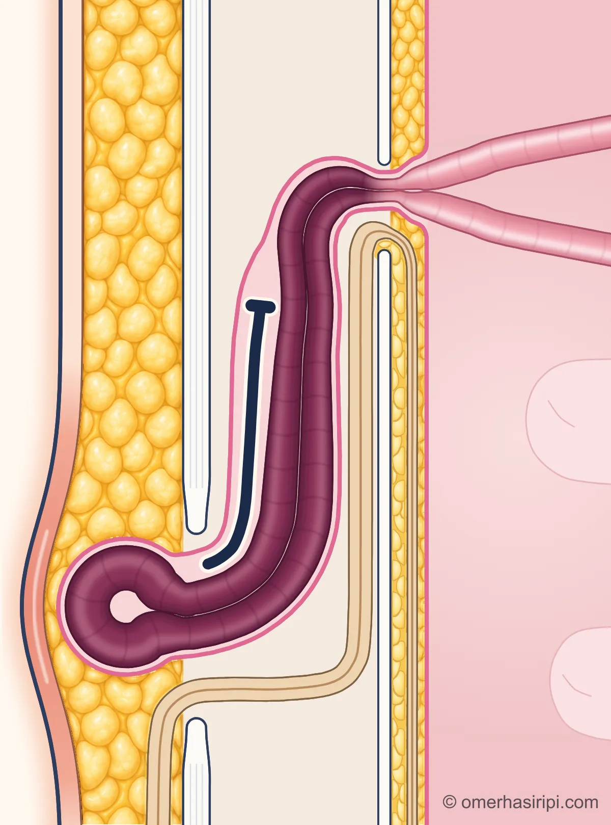
Schematic drawing: a section along the right inguinal canal of a man; the skin is on the left and the inside of the abdomen on the right, with the upper end towards the hip and the lower end towards the pubic bone. From the outside in, the drawing shows the skin, the fatty tissue beneath the skin, the tough white layer that forms the front wall of the canal, the canal itself, the thin back wall of the canal, a thin layer of fat and the abdominal lining as a pink line. The opening at the upper end of the back wall is the inner opening of the canal, and the opening at the lower end of the front wall is its outer opening. The spermatic cord enters the canal through the inner opening, runs down along the canal and comes out beneath the skin through the outer opening. A sac formed from the abdominal lining has entered the canal through the inner opening, run down on the skin side of the cord, come out beneath the skin through the outer opening and made the skin bulge. The sac and the bulge in the skin are larger and tense. A swollen, U-shaped pink loop of bowel fills the inside of the sac; its two limbs are squeezed into a narrow waist at the inner opening; inside the abdomen they separate and continue as pink bowel of normal thickness. A navy-blue arrow rising beside the sac stops at a short perpendicular line inside the canal.
Enlarge

Swipe the drawing right or left to see the details. You can use the arrow keys on your keyboard.

Schematic drawing: a section along the right inguinal canal of a man; the skin is on the left and the inside of the abdomen on the right, with the upper end towards the hip and the lower end towards the pubic bone. From the outside in, the drawing shows the skin, the fatty tissue beneath the skin, the tough white layer that forms the front wall of the canal, the canal itself, the thin back wall of the canal, a thin layer of fat and the abdominal lining as a pink line. The opening at the upper end of the back wall is the inner opening of the canal, and the opening at the lower end of the front wall is its outer opening. The spermatic cord enters the canal through the inner opening, runs down along the canal and comes out beneath the skin through the outer opening. A sac formed from the abdominal lining has entered the canal through the inner opening, run down on the skin side of the cord, come out beneath the skin through the outer opening and made the skin bulge. The sac and the bulge in the skin are larger and tense. A swollen, U-shaped pink loop of bowel fills the inside of the sac; its two limbs are squeezed into a narrow waist at the inner opening; inside the abdomen they separate and continue as pink bowel of normal thickness. A navy-blue arrow rising beside the sac stops at a short perpendicular line inside the canal.
  1. 1 Hernia sac (saccus herniae)
  2. 2 Small bowel trapped in the hernia (intestinum tenue)
  3. 3 Inner opening of the inguinal canal (anulus inguinalis profundus)
  4. 4 Cannot return inside
  5. 5 Outer opening of the inguinal canal (anulus inguinalis superficialis)
  6. 6 Spermatic cord (funiculus spermaticus)

If the contents become trapped in the sac and cannot go back into the abdomen, the hernia is trapped (incarceration). If your hernia normally goes back in easily but suddenly does not, see a doctor immediately.[1, 15, 16, 17]

Illustrative drawing, not to scale. Prepared for this site.

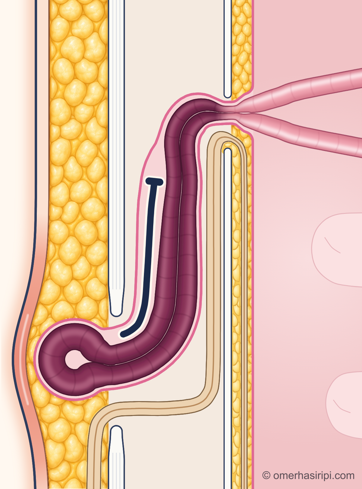
Schematic drawing: a section along the right inguinal canal of a man; the skin is on the left and the inside of the abdomen on the right, with the upper end towards the hip and the lower end towards the pubic bone. From the outside in, the drawing shows the skin, the fatty tissue beneath the skin, the tough white layer that forms the front wall of the canal, the canal itself, the thin back wall of the canal, a thin layer of fat and the abdominal lining as a pink line. The opening at the upper end of the back wall is the inner opening of the canal, and the opening at the lower end of the front wall is its outer opening. The spermatic cord enters the canal through the inner opening, runs down along the canal and comes out beneath the skin through the outer opening. A sac formed from the abdominal lining has entered the canal through the inner opening, run down on the skin side of the cord, come out beneath the skin through the outer opening and made the skin bulge. The appearance is the same as for the trapped hernia: a large, tense sac and a swollen loop of bowel whose two limbs are squeezed into a narrow waist at the inner opening; however, the loop inside the sac is dark purplish-red, the limbs on the abdominal side of the inner opening are normal pink bowel, and the skin over the bulge is red. A navy-blue arrow rising beside the sac stops at a short perpendicular line inside the canal.
Enlarge

Swipe the drawing right or left to see the details. You can use the arrow keys on your keyboard.

Schematic drawing: a section along the right inguinal canal of a man; the skin is on the left and the inside of the abdomen on the right, with the upper end towards the hip and the lower end towards the pubic bone. From the outside in, the drawing shows the skin, the fatty tissue beneath the skin, the tough white layer that forms the front wall of the canal, the canal itself, the thin back wall of the canal, a thin layer of fat and the abdominal lining as a pink line. The opening at the upper end of the back wall is the inner opening of the canal, and the opening at the lower end of the front wall is its outer opening. The spermatic cord enters the canal through the inner opening, runs down along the canal and comes out beneath the skin through the outer opening. A sac formed from the abdominal lining has entered the canal through the inner opening, run down on the skin side of the cord, come out beneath the skin through the outer opening and made the skin bulge. The appearance is the same as for the trapped hernia: a large, tense sac and a swollen loop of bowel whose two limbs are squeezed into a narrow waist at the inner opening; however, the loop inside the sac is dark purplish-red, the limbs on the abdominal side of the inner opening are normal pink bowel, and the skin over the bulge is red. A navy-blue arrow rising beside the sac stops at a short perpendicular line inside the canal.
  1. 1 Hernia sac (saccus herniae)
  2. 2 Small bowel with an impaired blood supply (intestinum tenue)
  3. 3 Inner opening of the inguinal canal (anulus inguinalis profundus)
  4. 4 Cannot return inside
  5. 5 Outer opening of the inguinal canal (anulus inguinalis superficialis)
  6. 6 Spermatic cord (funiculus spermaticus)

If the blood supply to the contents of the hernia is impaired, this is called a strangulated hernia (strangulation). If the hernia is suddenly much more painful than usual, red or larger, get medical help immediately. In the drawing, the bowel with an impaired blood supply is shown in a dark colour.[1, 15, 16, 17]

Illustrative drawing, not to scale. Prepared for this site.

What is a groin hernia and where does it occur?

In a groin hernia (inguinal hernia), contents of the abdomen push out through the inguinal canal[1]. They emerge through a weak spot in the abdominal wall around the line where the leg meets the abdomen[1]. What comes out may be fatty tissue or bowel[1].

A hernia in the same area may also emerge through the femoral canal[1]. This is called a thigh hernia (femoral hernia)[1]. Details are on the Femoral hernia page.

What causes a groin hernia?

The risk factors listed by the international guidelines are[1]:

  • A family history of groin hernia[1].
  • A previous hernia on the other side[1].
  • Being male and older age[1].
  • A disorder in the production of collagen (a connective tissue protein)[1].
  • Surgery to remove the prostate, especially complete removal by the open method[1].
  • Low body weight[1].

The evidence about the effects of heavy lifting, smoking and chronic constipation is weak or contradictory[1, 3].

What are the symptoms of a groin hernia?

The first sign is often a swelling in the groin[1]. There may be pain or a vague discomfort, but not always[1]. Pain and swelling may increase when you cough, sneeze, strain, lift a load or stand for a long time[1].

The swelling usually disappears when you lie down and relax[4].

Sometimes groin pain alone, without a swelling, may also be due to a hernia[1]. However, groin pain may have other causes[4]. It may also come from the hip, the area around the pubic bone, the muscles in this area or pressure on a nerve[4].

The diagnosis is usually made by examination[1]. Tests such as ultrasound or MRI are only needed if the diagnosis is uncertain[1]. Every woman with a hernia in the groin should also be checked for a femoral hernia[1].

In women, no examination or test can reliably distinguish between the two[1].

Which department treats a groin hernia?

Groin hernias are treated by the general surgery department[1]. If you suspect you have a groin hernia, see your family doctor or a surgeon[1]. Your family doctor can refer you for tests or treatment if necessary[5].

Is a groin hernia dangerous?

The contents of the hernia may suddenly become trapped and unable to return into the abdomen (trapped hernia)[1, 2]. If the blood supply to the trapped tissue is impaired, the tissue may begin to die (strangulated hernia)[1, 2]. A hernia that previously went back in but suddenly no longer does so is a potentially life-threatening emergency[2].

Although the evidence is weak, it has been reported that the likelihood of strangulation and the need to remove part of the bowel doubles every 24 hours after signs of trapping begin[2].

The risk of a groin hernia becoming trapped is less than 3% per year[1]. For hernias in the groin area, the risk of trapping and strangulation is higher in women, in femoral hernias and in people who have been admitted to hospital because of a hernia in the past year[1]. The risk of death with emergency surgery is 10–20 times higher than with planned surgery[1].

How does a groin hernia go away and what helps?

A groin hernia does not disappear without treatment[1]. It often grows over time and causes more pain[1]. A hernia can only be repaired with surgery[3].

According to the European Hernia Society's 2009 guidelines, a hernia truss (hernia belt) is now very rarely recommended[3].

When is groin hernia surgery necessary?

Surgery is recommended for a groin hernia that causes symptoms[1]. Watching and waiting is a safe option for men with few or no symptoms[1, 3]. The risk of an emergency problem is low in these men[1, 3].

However, most of these men eventually have surgery[1, 2]. The reason for surgery is most often the onset of pain[1, 2]. In studies, approximately 7 out of 10 people had surgery within 7–10 years[1, 2].

Complications, pain and discomfort were found to be similar in those who had surgery immediately and those who waited and had surgery later[2].

Outside pregnancy, watching and waiting is not recommended for women with a hernia in the groin area[1]. Femoral hernias are more common in women than in men[1]. The risk of strangulation is higher in women[1].

For this reason, outside pregnancy, timely surgery is recommended for women with a hernia in the groin area, even if there are no symptoms[1].

A swelling that appears in the groin during pregnancy is often not a hernia[1]. It is often an enlarged vein that goes away after childbirth[1]. It is appropriate to confirm the diagnosis with colour Doppler ultrasound, which also shows blood flow[1].

Even if a groin hernia develops during pregnancy, watching and waiting is recommended if there are no emergency signs (the evidence is weak)[1].

How is groin hernia surgery done and how long does it take?

Surgery is performed using an open or keyhole method[1]. In the open method, one incision is made in the groin[1]. In the keyhole (laparoscopic) method, a camera and fine instruments are used through one or several small incisions[1].

For most patients, reinforcing the place where the hernia emerges with a patch (mesh) is recommended[2]. The patch reduces the risk of the hernia coming back without increasing the risk of chronic pain[2]. Shrinking or movement of the patch is uncommon but can cause chronic pain[2].

A true allergy to the patch is extremely rare[2].
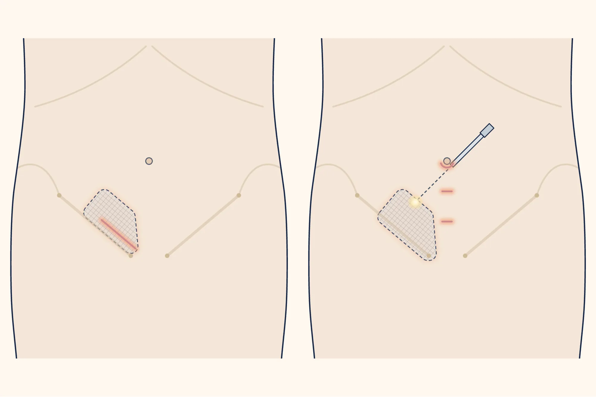
Two schematic drawings of the lower abdomen viewed from the front, side by side; the person's right is on the left of the picture. Both drawings show the same pale beige torso, faint arcs marking the lower edge of the rib cage and the upper edges of the hip bones, the belly button, and faint inguinal ligaments running downwards and inwards on both sides from the bony prominence at the front of the hip bone to the prominence of the pubic bone. Along the inner two-thirds of the inguinal ligament on the left of the picture is a light grey patch with rounded corners, a fine grid texture, a dashed outline and a faint peach glow, covering the place where the hernia emerges; the ligament can be seen through the patch. In the left drawing (open method), the lower edge of the patch is above the ligament; there is one straight incision just above and parallel to the inner half of the ligament, with its inner end finishing just above the pubic bone prominence. In the right drawing (keyhole method), the patch is in the same place, with its lower edge extending slightly below the ligament; there are three small incisions on the midline: a slightly larger one at the lower edge of the belly button and two small incisions between the belly button and the pubic bone. A thin camera rod enters at an angle through the incision at the belly button; its outer part is shown as a line and its inner part as a dashed line descending towards the right groin. The dashed line ends in a small, soft light near the patch. There is no incision or patch in the left groin.
Enlarge

Swipe the drawing right or left to see the details. You can use the arrow keys on your keyboard.
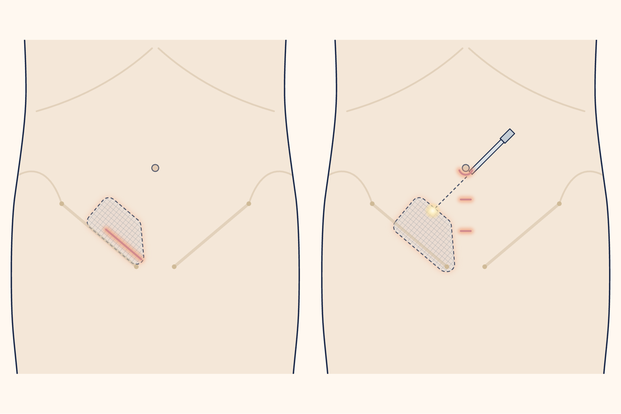
Two schematic drawings of the lower abdomen viewed from the front, side by side; the person's right is on the left of the picture. Both drawings show the same pale beige torso, faint arcs marking the lower edge of the rib cage and the upper edges of the hip bones, the belly button, and faint inguinal ligaments running downwards and inwards on both sides from the bony prominence at the front of the hip bone to the prominence of the pubic bone. Along the inner two-thirds of the inguinal ligament on the left of the picture is a light grey patch with rounded corners, a fine grid texture, a dashed outline and a faint peach glow, covering the place where the hernia emerges; the ligament can be seen through the patch. In the left drawing (open method), the lower edge of the patch is above the ligament; there is one straight incision just above and parallel to the inner half of the ligament, with its inner end finishing just above the pubic bone prominence. In the right drawing (keyhole method), the patch is in the same place, with its lower edge extending slightly below the ligament; there are three small incisions on the midline: a slightly larger one at the lower edge of the belly button and two small incisions between the belly button and the pubic bone. A thin camera rod enters at an angle through the incision at the belly button; its outer part is shown as a line and its inner part as a dashed line descending towards the right groin. The dashed line ends in a small, soft light near the patch. There is no incision or patch in the left groin.
  1. 1 Open method: one incision in the groin
  2. 2 Keyhole (laparoscopic) method: small incisions
  3. 3 Camera
  4. 4 Patch (mesh)
  5. 5 Inguinal ligament: the ligament running from the bony prominence at the front of the hip bone to the pubic bone
  6. 6 Belly button
  7. 7 Person's right side
Description of the drawing

Open method on the left, keyhole (laparoscopic) method on the right; the hernia is in the person's right groin. In the open method, one incision is made in the groin; in the keyhole method, a camera and fine instruments are used through several small incisions. The location and number of incisions may vary; the drawing shows one commonly used arrangement. The grid-patterned area shows the position in the abdominal wall of the patch that strengthens the place where the hernia emerges, not its depth.[6, 8]

Results may vary from person to person with any surgical or interventional procedure. You are advised to discuss the procedure in detail with your doctor beforehand.

Illustrative drawing, not to scale. Source: Cancer Research UK / Wikimedia Commons, CC BY-SA 4.0. Only the outline of the torso was taken from the appendix drawing adapted from this source; everything else was newly drawn for this site. This adaptation is also licensed under CC BY-SA 4.0.

For a first-time hernia on one side, the keyhole method is recommended if a surgeon experienced in the method and adequate facilities are available[2]. According to the guidelines, there is less pain after surgery and less chronic pain with this method[2]. Recovery is faster[2].

For some patient and hernia characteristics, the open method is the first choice[2]. In the long term, the rate of hernia recurrence is no different between the two methods[2]. The keyhole method is also recommended for hernias on both sides[1].

In women, this method also helps prevent a femoral hernia from being missed[1].

Open surgery is performed under a regional anaesthetic (for example, a spinal anaesthetic given in the back) or a general anaesthetic[1, 2]. Keyhole surgery is usually performed under a general anaesthetic[1, 2]. The options are discussed with you[1, 2].

Surgery on one side usually takes 45 minutes to one hour[3]. Surgery on both sides may take longer; the additional time depends on the method used[1].

Problems caused by bleeding after surgery are more common in people taking blood thinners or medicines that prevent clotting[1]. Do not stop these medicines on your own. Ask the doctor who prescribed them what to do.

Open method, TAPP and TEP: what are the differences?

The Lichtenstein operation is the benchmark and most commonly used method for open repair with a patch[1]. Entry is through the incision in the groin[3]. The inguinal canal is reached by opening the flat tendon layer of the external oblique muscle (aponeurosis)[3].

The patch is stitched to the back wall of the canal, beneath this layer[3].

There are two commonly used keyhole methods: TAPP and TEP. The abdominal lining is the thin membrane that lines the inside of the abdominal cavity[1]. In both methods, the patch is placed in the space in front of the abdominal lining[1].

The difference is how this space is reached[1]. TAPP stands for transabdominal preperitoneal: through the abdominal cavity to the space in front of the abdominal lining[1]. In TAPP, the abdominal cavity is first entered with a camera[1].

This allows the surgeon to see at the start of the operation whether there is also a hernia in the other groin[1]. The abdominal lining is opened, the patch is placed, and the incision in the lining is carefully closed[1, 13]. TEP stands for totally extraperitoneal: entirely outside the abdominal lining[1].

In TEP, the abdominal cavity is not entered[1]. The same space is reached directly[1]. There is no need to open and close the abdominal lining[1].
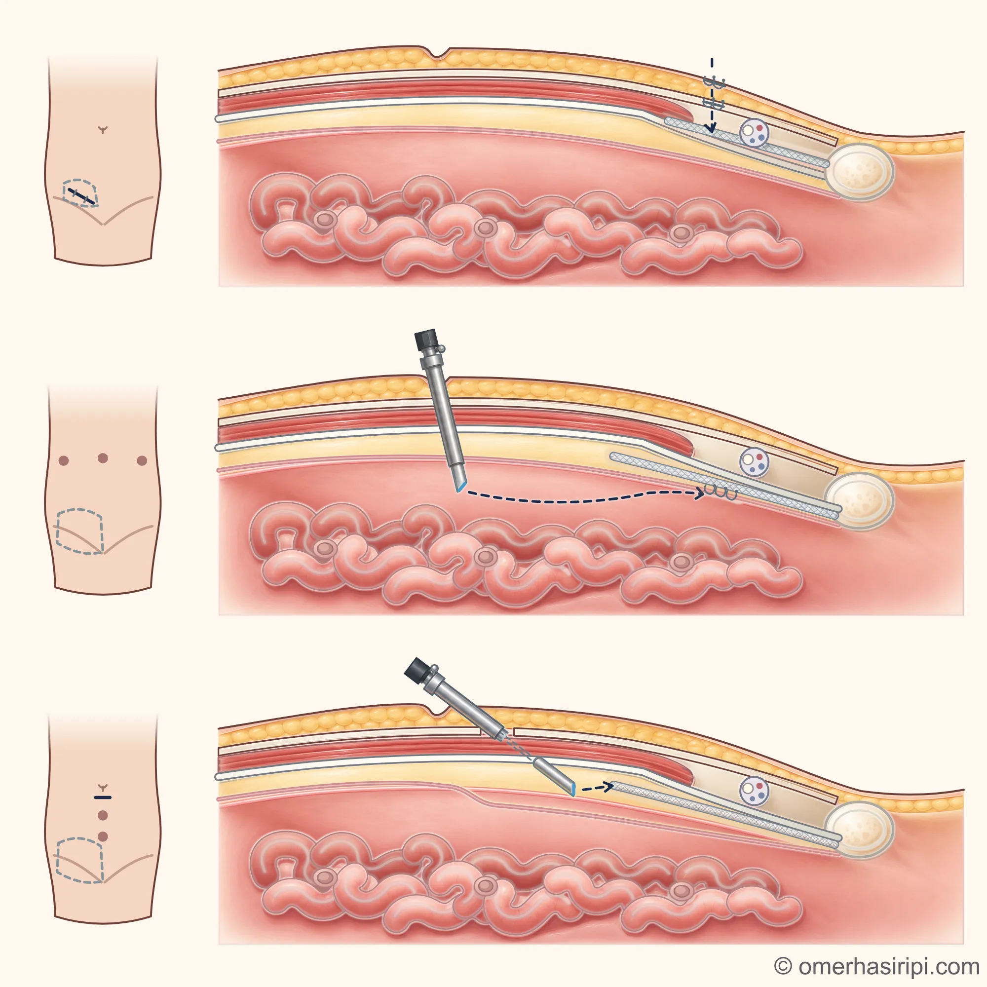
Three panels stacked vertically. In each panel, a man's lower abdomen is shown from the front on the left, and the same area is shown as a schematic side section on the right: the patient is lying on his back, with his head to the left; the abdominal wall with the belly button is at the top, the abdominal cavity containing the bowel is inside, and the groin and pubic bone are at the lower right. At the top (open method), there is one incision in the groin; the patch is on the back wall of the inguinal canal; the incisions in the skin and the tendon layer have been closed. In the middle (TAPP), three small incisions are shown as an example; a camera entering through one of the incisions is in the abdominal cavity; a larger patch is behind the transversalis fascia and in front of the abdominal lining; the incision in the abdominal lining has been closed. At the bottom (TEP), one incision below the belly button and two small incisions for instruments are shown as an example; the camera enters through the front sheath of the rectus abdominis muscle and extends into the space in front of the abdominal lining; the same patch is in the same place, and the abdominal lining has not been opened.
Enlarge

Swipe the drawing right or left to see the details. You can use the arrow keys on your keyboard.
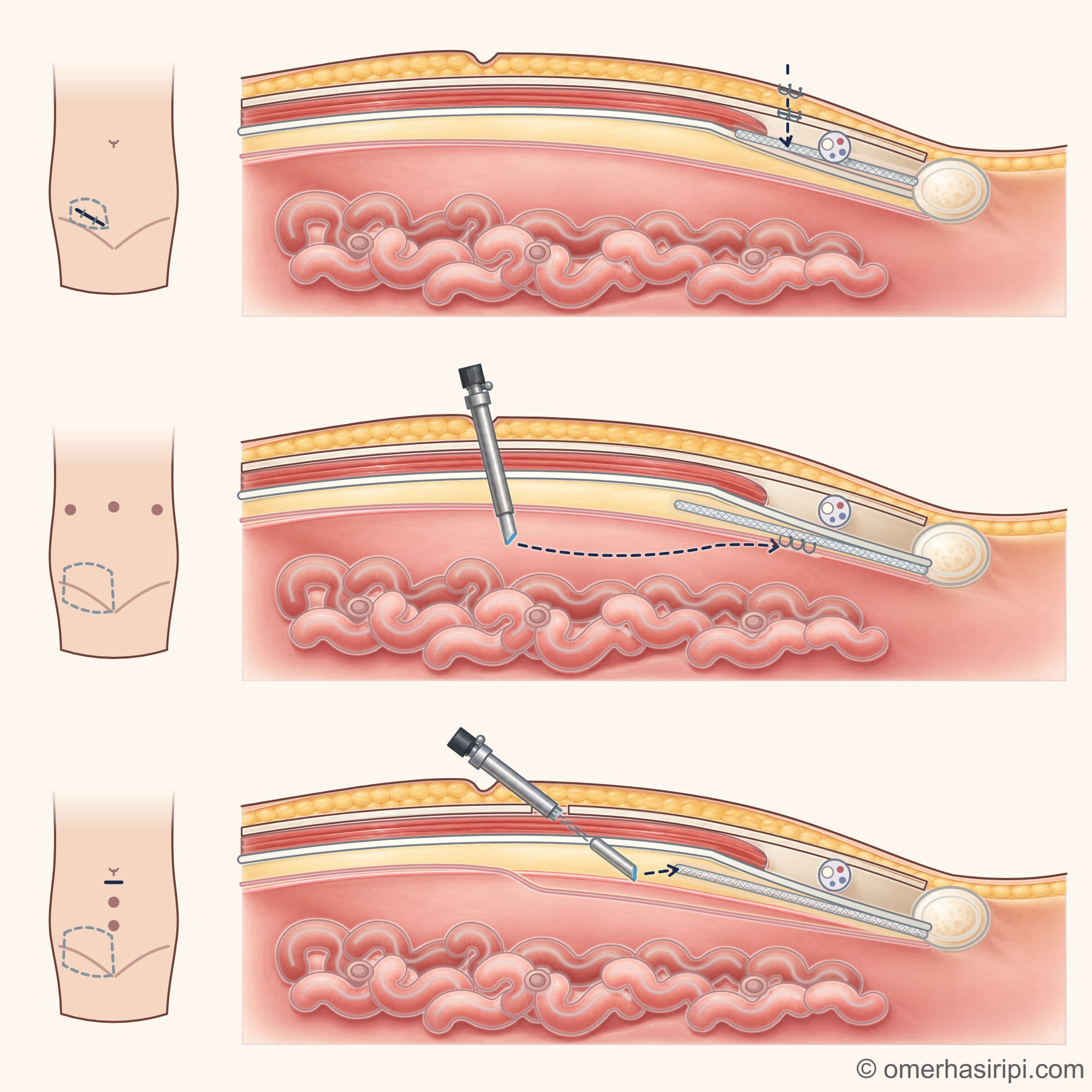
Three panels stacked vertically. In each panel, a man's lower abdomen is shown from the front on the left, and the same area is shown as a schematic side section on the right: the patient is lying on his back, with his head to the left; the abdominal wall with the belly button is at the top, the abdominal cavity containing the bowel is inside, and the groin and pubic bone are at the lower right. At the top (open method), there is one incision in the groin; the patch is on the back wall of the inguinal canal; the incisions in the skin and the tendon layer have been closed. In the middle (TAPP), three small incisions are shown as an example; a camera entering through one of the incisions is in the abdominal cavity; a larger patch is behind the transversalis fascia and in front of the abdominal lining; the incision in the abdominal lining has been closed. At the bottom (TEP), one incision below the belly button and two small incisions for instruments are shown as an example; the camera enters through the front sheath of the rectus abdominis muscle and extends into the space in front of the abdominal lining; the same patch is in the same place, and the abdominal lining has not been opened.
  1. 1 Open method (Lichtenstein): through the incision in the groin
  2. 2 TAPP: entry through the abdominal cavity
  3. 3 TEP: the abdominal cavity is not entered
  4. 4 Front view: location of the incisions; the dashed line shows the location of the patch
  5. 5 Belly button
  6. 6 Skin and underlying fatty tissue
  7. 7 Abdominal wall muscle and its front sheath
  8. 8 Flat tendon layer of the external oblique muscle (aponeurosis)
  9. 9 Spermatic cord
  10. 10 Back wall of the inguinal canal (transversalis fascia)
  11. 11 Pubic bone
  12. 12 Abdominal lining (peritoneum)
  13. 13 Abdominal cavity and bowel
  14. 14 Patch: on the back wall of the inguinal canal
  15. 15 Patch: behind the transversalis fascia, in front of the abdominal lining
  16. 16 The incision in the abdominal lining is closed after the patch is placed
  17. 17 Patch: in the same space, without opening the abdominal lining
Description of the drawing

In each panel, the lower abdomen is shown from the front on the left: the incisions are shown, and the dashed line marks the position of the patch. On the right, the same area is shown as a simplified side section, as if viewed from the side while the patient is lying on his back: the head end is on the left and the front of the abdomen is at the top. In the groin, the back wall of the inguinal canal is formed mainly by a thin membrane (the transversalis fascia). At the top, the open method (Lichtenstein): the surgeon enters through the incision in the groin; the patch is stitched to the back wall of the inguinal canal. In the middle, TAPP: the abdominal cavity is entered through a small incision, the abdominal lining is opened, the patch is placed behind the transversalis fascia, in the space in front of the abdominal lining, and the incision in the lining is closed. At the bottom, TEP: the surgeon enters the front sheath of the rectus abdominis muscle through an incision just below the belly button and opens the same space without entering the abdominal cavity; there is no need to open the abdominal lining, although it may be torn accidentally. The location and number of incisions in keyhole methods may vary; the drawing shows an example arrangement. The drawing shows a man.[1, 3, 13]

Results may vary from person to person with any surgical or interventional procedure. You are advised to discuss the procedure in detail with your doctor beforehand.

Illustrative drawing, not to scale. Prepared for this site.

Because the abdominal cavity is not entered, injury to internal organs such as the bowel has been reported less often with TEP than with TAPP, although this difference has not been found to be certain in every study[1]. Hernias developing later at the small incisions where the camera and instruments enter have also been reported less often with TEP than with TAPP[1, 3]. These problems are uncommon with both methods[1].

According to the international guidelines, apart from these differences, TAPP and TEP have similar operation times and overall complication risks[1]. Rates of pain after surgery, chronic pain and hernia recurrence are also similar[1]. The guidelines consider both methods suitable for treating groin hernias[1].

The guidelines consider the surgical team's experience important when choosing a method[1]. Blood vessel injury is a very rare but serious complication with both methods[1]. Injury to blood vessels while entering and working in the space in front of the abdominal lining has been reported slightly more often with TEP than with TAPP[1].

Completing the operation by switching to another method has been reported slightly more often with TEP than with TAPP[1, 13]. For example, if the abdominal lining is accidentally torn, the repair may be completed with TAPP[1, 13]. Learning keyhole methods, especially TEP, takes longer than learning the open method[2].

Rare but severe complications have been seen more often while the surgeon is learning the method[2].

In general surgical practice, the guidelines consider some situations relative contraindications to the keyhole method[2]:

  • Previous prostate surgery[2].
  • Having received radiotherapy to the pelvis (the area inside the pelvic bones)[2].
  • Previous lower abdominal (pelvic) surgery[2].
  • A hernia that descends into the scrotum[2].
  • A lack of experience or facilities for the keyhole method[2].

In these situations, open repair from the front is a better option[2]. The method should be chosen according to the patient, the type of hernia and the surgeon's experience[2]. If the hernia has come back at a previously operated site, the method is chosen according to the previous operation[1].

  • If it was repaired from the front with a patch (for example, using the Lichtenstein method), repair from behind (for example, using TAPP or TEP) is recommended[1].
  • If it was repaired from behind, repair from the front is recommended[1].
  • If the previous repair was performed without a patch, using stitches alone, either approach can be used[1].

If a hernia that has been repaired both from the front and from behind comes back, it is recommended that treatment be left to a surgeon who specialises in hernia surgery[1].

Is groin hernia surgery dangerous?

Groin hernia surgery is successful in most patients[1]. The rates below come from studies brought together in the guidelines. They are not any individual doctor's own results.

A large review found a complication rate of approximately 3% around the time of surgery[1, 2]. The same review found a wound infection rate of 5 per 1,000[1, 2]. Higher rates have also been reported in other studies[1, 2].

  • Inability to urinate: from less than 1% to 20%[1]. This is most common after a general or regional anaesthetic[1].
  • Moderate or severe sexual function problems: approximately 5–6%[1]. Impaired testicular function or fertility: less than 1%[1].
  • Here, chronic pain means groin pain that lasts at least 3 months after surgery, is at least moderate, causes discomfort and affects daily activities[1]. It occurs in approximately 1 in 10 people and decreases over time[1]. Pain that limits daily life or work: 0.5–6%[1].
  • Injury to the bowel, bladder or blood vessels is rare[1]. It is slightly more common with the keyhole method[1].

Death within 30 days after surgery is very rare and is mostly associated with emergency surgery or other conditions[1]. The hernia may come back in some people[1]. Reported rates vary greatly according to the measurement method, length of follow-up and centre[1, 2].

How long does it take to recover from groin hernia surgery?

Step by step after surgery

The following are approximate times and recommendations from the sources for after planned surgery that has gone smoothly[1]. They are not dates for full recovery[1]. Recovery usually takes up to 4–6 weeks[6]. Sometimes it may take longer[6]. Your surgeon will tell you the plan that is right for you[3].

  1. First few days

    Move around and support the operation site

    Walk as much as you can and gradually increase your activity[6]. Support the operation site when you cough, sneeze or strain[1].

  2. 1–2 weeks

    Pain relief

    Some pain or discomfort is normal[1]. Most patients are expected to feel relief within 1–2 weeks[1].

  3. Depending on the person and the job

    Daily activities and returning to work

    The sources' recommendations on returning to work are in the “For doctors and those interested” section at the end of the page.

  4. Depending on the person

    Heavy lifting and contact sports

    When you return to lifting depends on your operation and personal circumstances[3]. Your surgeon will advise you[3]. The different timeframes given by the sources are in the “For doctors and those interested” section at the end of the page.

Details: When should you see a doctor after surgery?

After groin hernia surgery: when should you see a doctor?

A collection of blood (haematoma) that causes bruising and swelling may develop at the operation site[1]. Most go away on their own within 2–4 weeks[1]. See a doctor urgently if it causes severe pain or significant swelling, or if the skin over it turns black[1].

Bruising or swelling around the genitals is normal for a few weeks after surgery[6]. Get medical help without delay if any of the following occur[6]:

  • pus, pain, swelling, warmth or redness in the wound, or heavy bleeding from the wound[6],
  • a high temperature, feeling cold or shivering, increasing abdominal swelling or pain[6],
  • nausea or vomiting that continues 24 hours after surgery[6],
  • throbbing or cramp-like pain in the leg, new or worsening shortness of breath that is not severe, coughing up a few small spots or streaks of blood[6].

If you have difficulty urinating, contact your surgeon immediately[12].

Severe shortness of breath, pain in the chest or upper back, coughing up more than a few small spots or streaks of blood, or a very fast, very slow or irregular heartbeat may be a sign of a blood clot in the lung: call 112 or go to the nearest emergency department[6]. Do not drive yourself to the emergency department[6].

If the signs above are absent, contact your surgeon if pain does not go away with the recommended medicines or if it is moderate, severe or long-lasting[1]. Also contact your surgeon if the groin symptoms you had before surgery return: the hernia may have come back[1]. If the signs above are absent, call your surgeon if you have nausea or vomiting after surgery, cannot eat or drink fluids, or develop a cough[12].

How can you prepare for a groin hernia examination as an adult?

  • Make a note of when you first noticed the swelling or pain[11]. Describe any changes you have noticed while standing, coughing or doing daily activities[11, 12].
  • You can make a short list of the topics you particularly want to discuss at the examination[10].
  • Tell the doctor if you have had hernia or abdominal surgery before[10, 12].
  • Prepare a list of your prescription and non-prescription medicines, vitamins and supplements[11]. Make a note of your known allergies[10]. Also tell the doctor about any other conditions you have and any difficulty urinating you have experienced before[10, 12].
  • Prioritise two or three questions[11]. Example questions:

    • “What should the next step be for me?”[11]
    • “If surgery is recommended, which method is suitable and why?”[11, 12]
    • “What plan should I make for my own work and daily activities?”[12]

    Write down the answers and the next step you agree on[11].

Sources

17
  1. HerniaSurge Group. International guidelines for groin hernia management. Hernia. 2018;22(1):1–165. doi: 10.1007/s10029-017-1668-x. PubMed: 29330835.
  2. Stabilini C, van Veenendaal N, Aasvang E, Agresta F, Aufenacker T, Berrevoet F, et al. Update of the international HerniaSurge guidelines for groin hernia management. BJS Open. 2023;7(5):zrad080. doi: 10.1093/bjsopen/zrad080. PubMed: 37862616.
  3. Simons MP, Aufenacker T, Bay-Nielsen M, Bouillot JL, Campanelli G, Conze J, et al. European Hernia Society guidelines on the treatment of inguinal hernia in adult patients. Hernia. 2009;13(4):343–403. doi: 10.1007/s10029-009-0529-7. PubMed: 19636493.
  4. Kirk M, Henriksen NA, Christoffersen MW, Andresen K, Dorfelt A, Krogsgaard M, et al. Management of Adult Groin Hernia in Denmark: a National Updated Consensus. J Abdom Wall Surg. 2025;4:14904. (Danish national consensus report; by its own description, it is not an official guideline.) doi: 10.3389/jaws.2025.14904. PubMed: 40860237.
  5. NHS (United Kingdom National Health Service). Hernia (hernia): patient information page. Last reviewed: 19 May 2026. Text: www.nhs.uk.
  6. NHS (United Kingdom National Health Service). Inguinal hernia repair (groin hernia surgery): patient information page. Last reviewed: 28 February 2025. Text: www.nhs.uk.
  7. NHS (United Kingdom National Health Service). Sudden confusion (delirium) (sudden confusion): patient information page. Last reviewed: 28 May 2024. Text: www.nhs.uk.
  8. Nawaz Y, Barvalia M, Rana G, Khakwani MZ, Azim K, et al. Poorly recognized age-related downward deviation of the inguinal ligament. SAGE Open Med. 2016;4:2050312116675565. (Full text: bony attachments of the inguinal ligament; accessed 4 October 2026.) doi: 10.1177/2050312116675565. PubMed: 27826446.
  9. Chiu MK, Hadied MO, Klochko C, van Holsbeeck MT. Comparison of patient characteristics and treatment approaches for femoral and inguinal hernias utilizing dynamic ultrasound at a single institution. Hernia. 2023;27:1245–1252. (Full text: Figure 3, relationship of the femoral hernia to the blood vessels; accessed 4 October 2026.) doi: 10.1007/s10029-023-02810-2. PubMed: 37253821.
  10. NHS — Outpatients and day patients. Last reviewed 21 April 2023; next review 21 April 2026 (overdue at the time of access). Accessed: 25 October 2026. Text: www.nhs.uk.
  11. NHS — What to ask your doctor or other healthcare professional. Last reviewed 12 January 2023; next review 12 January 2026 (overdue at the time of access). Accessed: 25 October 2026. Text: www.nhs.uk.
  12. SAGES — Inguinal Hernia Repair Surgery: Patient Information. Revised 6 May 2026. Accessed: 25 October 2026. Text: www.sages.org.
  13. Bittner R, Montgomery MA, Arregui E, Bansal V, Bingener J, Bisgaard T, et al. Update of guidelines on laparoscopic (TAPP) and endoscopic (TEP) treatment of inguinal hernia (International Endohernia Society). Surg Endosc. 2015;29(2):289–321. (Full text reviewed: 28 October 2026.) doi: 10.1007/s00464-014-3917-8. PubMed: 25398194.
  14. OpenStax. Anatomy and Physiology 2e. 2022. Section 23.1: Overview of the Digestive System; full text accessed 28 October 2026. Text: openstax.org.
  15. OpenStax. Anatomy and Physiology 2e. 2022. Section 27.1: Anatomy and Physiology of the Testicular Reproductive System; full text accessed 28 October 2026. Text: openstax.org.
  16. Yoong P, Duffy S, Marshall TJ. The inguinal and femoral canals: a practical step-by-step approach to accurate sonographic assessment. Indian J Radiol Imaging. 2013;23(4):391–395. (Full text: the two openings of the inguinal canal, the course of the hernia along the canal, fat or bowel in the sac; accessed 28 October 2026.) doi: 10.4103/0971-3026.125586. PubMed: 24604947.
  17. Yasukawa D, Aisu Y, Hori T. Crucial anatomy and technical cues for laparoscopic transabdominal preperitoneal repair: advanced manipulation for groin hernias in adults. World J Gastrointest Surg. 2020;12(7):307–325. (Full text: course of the sperm duct outside the abdominal lining; accessed 28 October 2026.) doi: 10.4240/wjgs.v12.i7.307. PubMed: 32821340.

Medical review of the Turkish original: Op. Dr. Ömer Seyfullah Hasıripi, . This page is a translation checked by AI reviewers; the Turkish version prevails.

This page is for general information; it does not replace an examination. See a doctor about your symptoms.