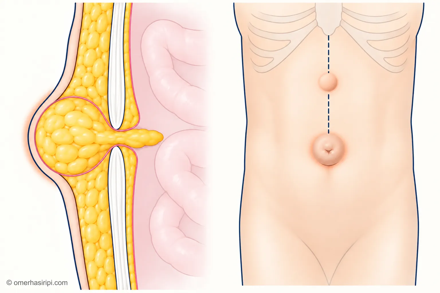
What is an umbilical hernia?
A hernia is when the contents of the abdomen protrude through an opening in the abdominal wall[8]. In an umbilical hernia, this opening is at the belly button: part of the bowel or the surrounding fatty tissue is pushed out through it and forms a bulge at the belly button[8, 9]. The opening is usually small (1–2 cm) and the hernia contains only fatty tissue; but bowel or other organs may also be present[8]. A hernia that emerges above the belly button, on the midline of the abdomen, is called an epigastric hernia[3, 8].
What causes an umbilical hernia?
In adults, conditions such as excess weight (obesity), more than one pregnancy, or liver disease that causes fluid to build up in the abdomen (ascites) can cause an umbilical hernia[9].
What are the symptoms of an umbilical hernia?
Most people with an umbilical hernia have very few symptoms or none at all; a bulge or lump is usually noticed around the belly button[8]. The bulge is more noticeable when standing; it may become smaller or disappear when you lie down[8]. The hernia is usually painless, but in adults it can sometimes cause discomfort when coughing or straining[9]. As the hernia grows, discomfort may appear or increase; this can take months or years[8].
The diagnosis is usually made by examination; if the examination is not clear, an ultrasound or a computed tomography (CT) scan may be needed, and for a large hernia, a CT scan may be needed to plan the surgery[8]. If your hernia causes no symptoms at all, you do not need to rush to see a doctor[8]. If the bulge at the belly button is painful or hard, is growing or does not go away, see a doctor, even if it is not an emergency[9]. If a hernia that previously went back in no longer does so, this is an emergency: see the “When is it an emergency?” section below[8].
A bulge between the breastbone and the belly button, most noticeable when doing sit-ups, may be a widening of the midline between the two abdominal muscles (rectus diastasis)[5, 8]. It is more common in women who have given birth and in middle-aged men[8]. Rectus diastasis is not a hernia and carries no risk of trapping or strangulation[4, 5]; but it can occur alongside an umbilical hernia[8].
When does an umbilical hernia become dangerous?
The contents of the hernia can become trapped outside (trapping, or incarceration); if the blood supply to the contents becomes insufficient, this is called a strangulated hernia (strangulation) and urgent treatment is needed[8]. Strangulation can lead to severe infection (sepsis) and tissue death (gangrene); surgery is needed as soon as possible[9]. These problems can sometimes occur in adults[9]. If fluid is building up in your abdomen because of scarring of the liver (cirrhosis), waiting without surgery can be risky; planned surgery may be considered if the fluid is under control and liver function is preserved[7]. In this situation, if abdominal pain starts or fluid leaks from the belly button, go to the emergency department immediately[7].
Enlarge
Swipe the drawing right or left to see the details. You can use the arrow keys on your keyboard.
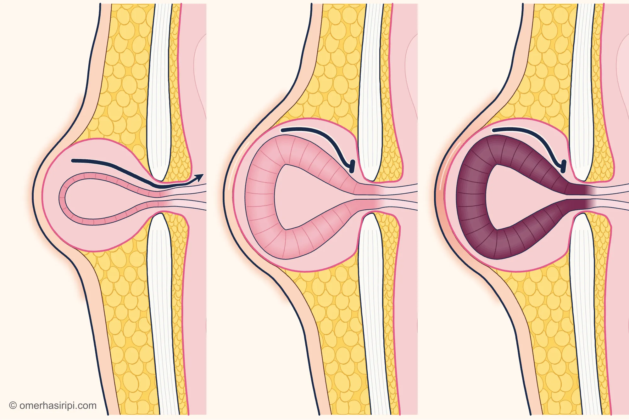
- 1 Hernia that can return inside the abdomen
- 2 Small bowel trapped in the hernia
- 3 Small bowel with an impaired blood supply
- 4 Opening in the abdominal wall
- 5 Bowel pushed outwards
- 6 Can return inside
- 7 Cannot return inside
- 8 Connective tissue supporting the abdominal wall
- 9 Hernia sac
From left to right: When you lie down, the hernia may become smaller or return inside the abdomen. If the contents become trapped outside the opening and cannot return, the hernia is trapped (incarceration); if a hernia that previously went back in no longer does so, urgent assessment is needed. If the blood supply to the trapped contents becomes impaired, this is called a strangulated hernia (strangulation) and urgent treatment is needed.[12, 14]
Illustrative drawing, not to scale. Prepared for this site.
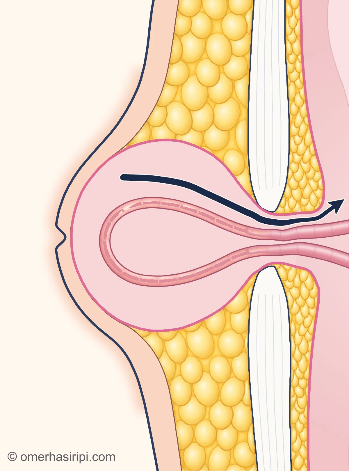
Enlarge
Swipe the drawing right or left to see the details. You can use the arrow keys on your keyboard.
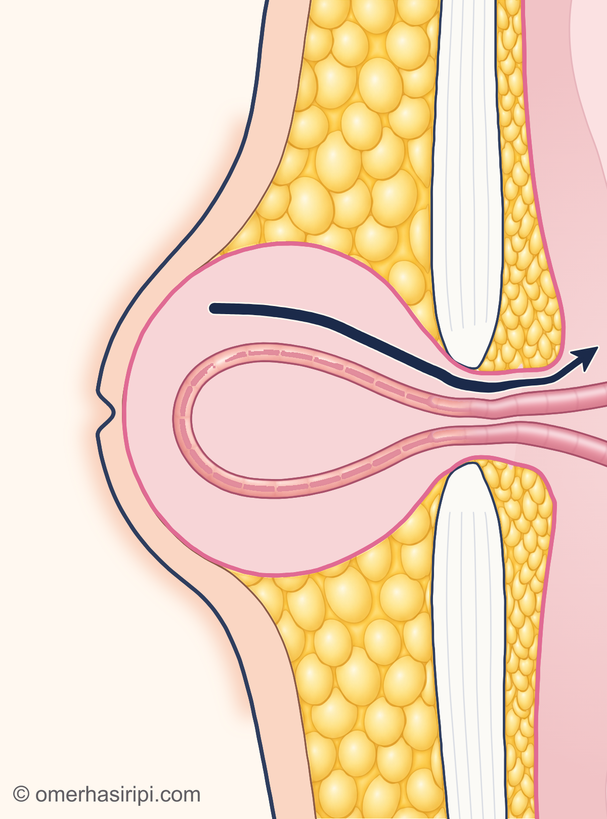
- 1 Hernia sac (saccus herniae)
- 2 Small bowel pushed outwards (intestinum tenue)
- 3 Opening in the abdominal wall (anulus umbilicalis)
- 4 Can return inside
- 5 Firm layer where the muscle sheaths meet in the middle (linea alba)
Description of the drawing
The hernia may also contain bowel. When you lie down, the hernia may become smaller or return inside the abdomen.[8, 12]
Illustrative drawing, not to scale. Prepared for this site.
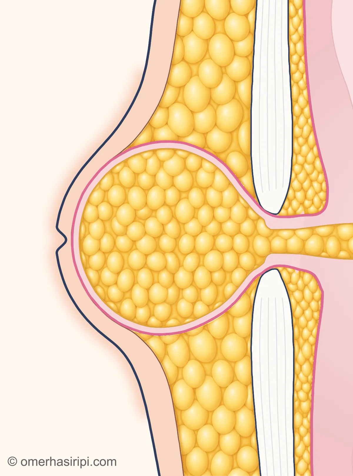
Enlarge
Swipe the drawing right or left to see the details. You can use the arrow keys on your keyboard.
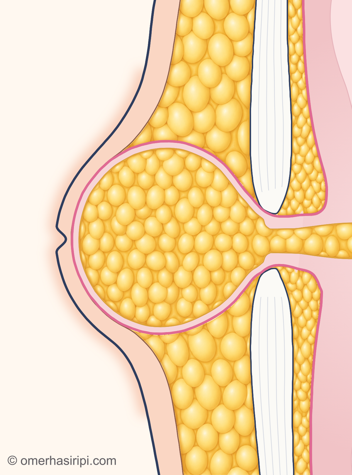
- 1 Hernia sac (saccus herniae)
- 2 Fatty apron inside the abdomen (omentum majus)
- 3 Opening in the abdominal wall (anulus umbilicalis)
- 4 Fat tissue beneath the skin
- 5 Firm layer where the muscle sheaths meet in the middle (linea alba)
Description of the drawing
In most umbilical hernias, the hernia contains only fatty tissue. In the drawing, this fat is a part of the fatty apron inside the abdomen (omentum) that has been pushed out through the opening.[6, 8, 15]
Illustrative drawing, not to scale. Prepared for this site.
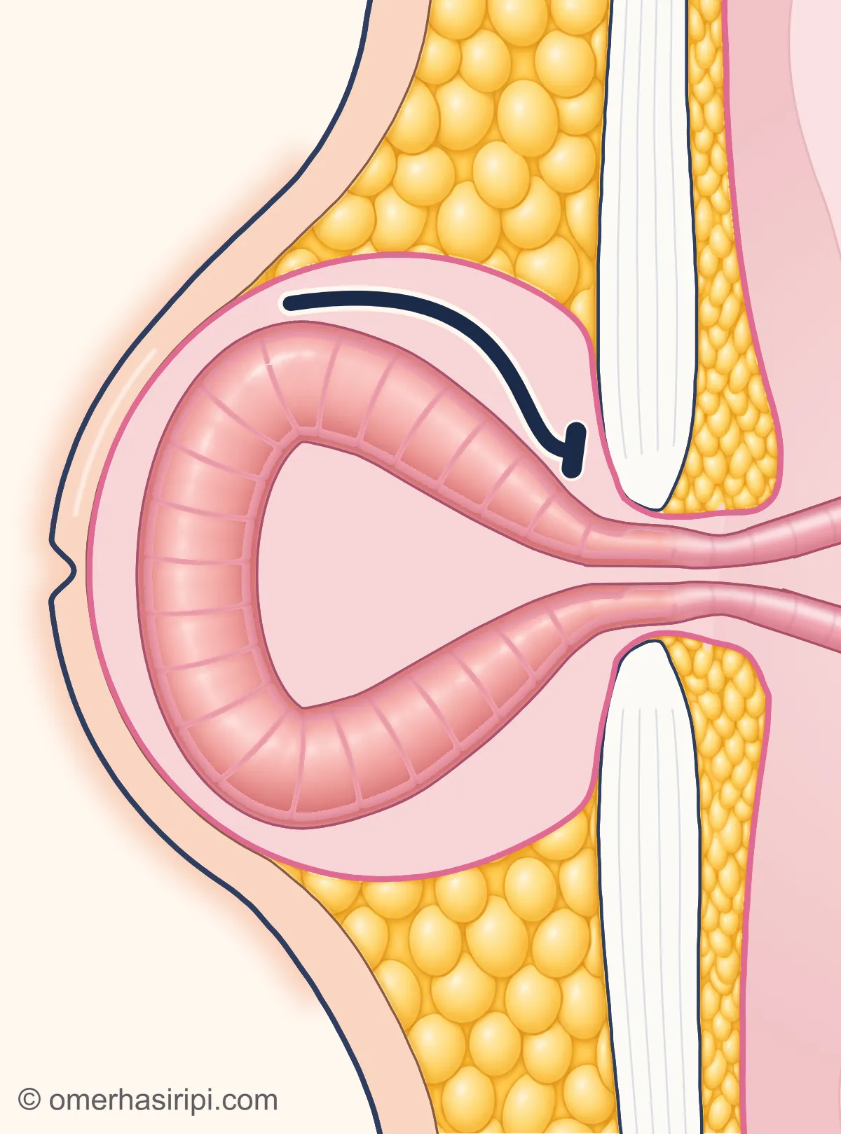
Enlarge
Swipe the drawing right or left to see the details. You can use the arrow keys on your keyboard.
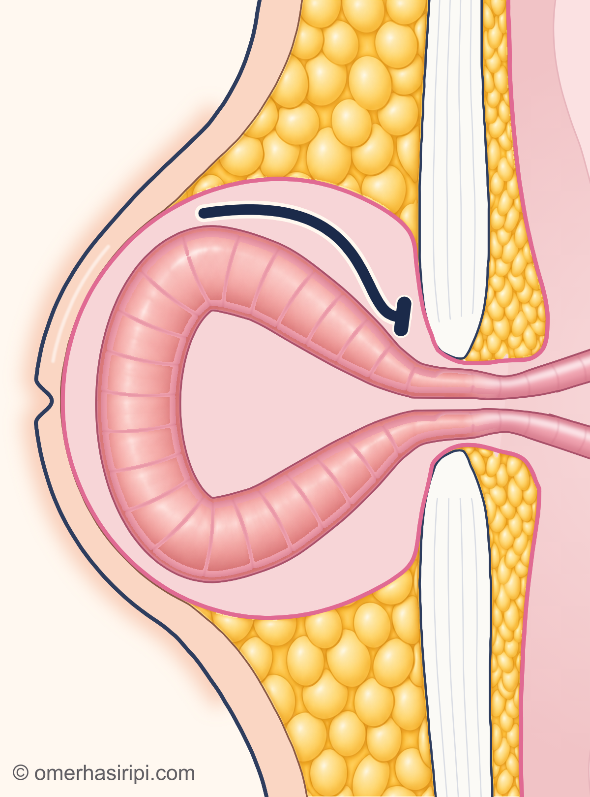
- 1 Hernia sac (saccus herniae)
- 2 Small bowel trapped in the hernia (intestinum tenue)
- 3 Opening in the abdominal wall (anulus umbilicalis)
- 4 Cannot return inside
- 5 Firm layer where the muscle sheaths meet in the middle (linea alba)
If the contents become trapped outside the opening and cannot return, the hernia is trapped (incarceration). If a hernia that previously went back in no longer does so, this is an emergency; get surgical help immediately.[8, 12]
Illustrative drawing, not to scale. Prepared for this site.
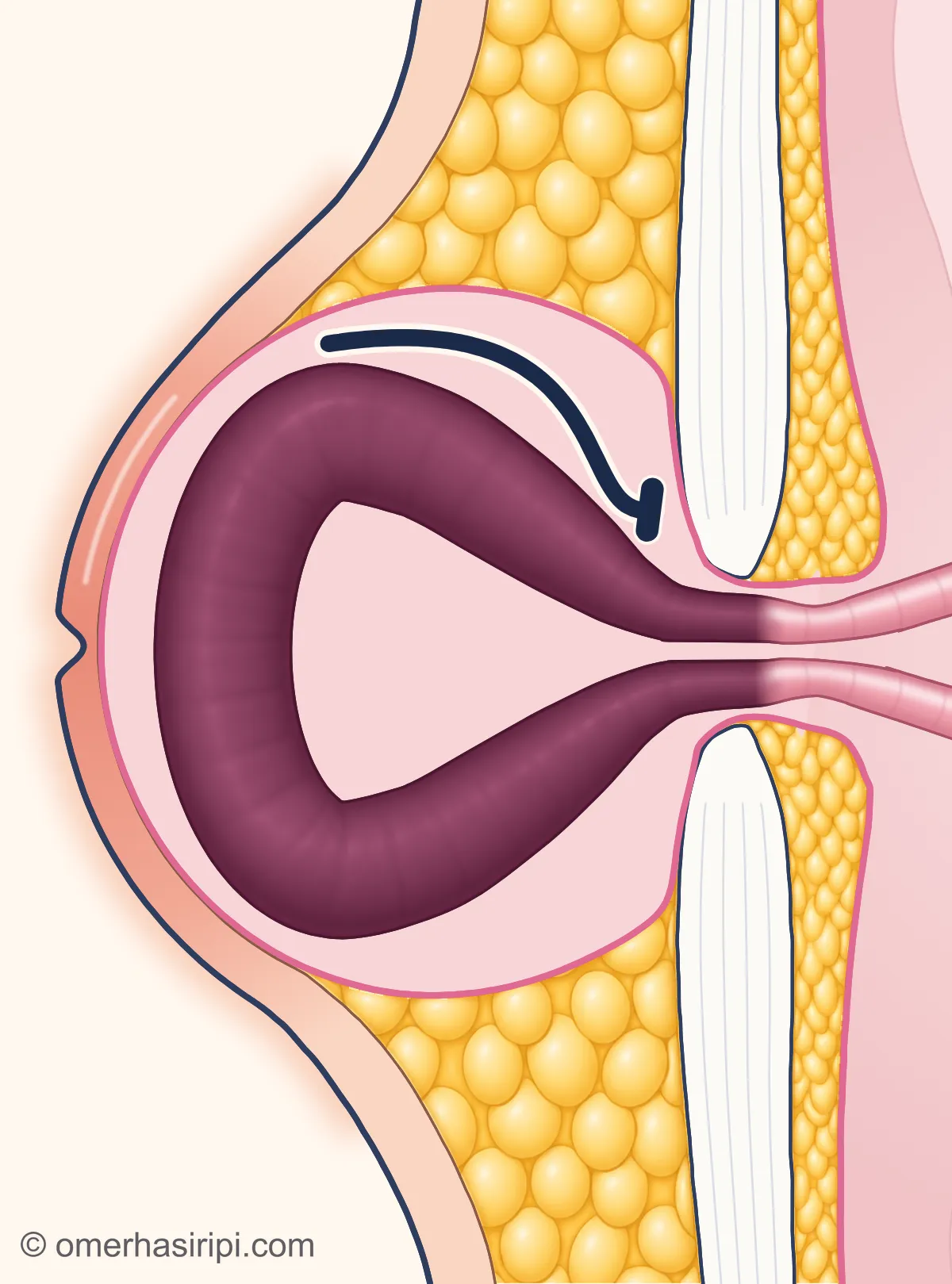
Enlarge
Swipe the drawing right or left to see the details. You can use the arrow keys on your keyboard.
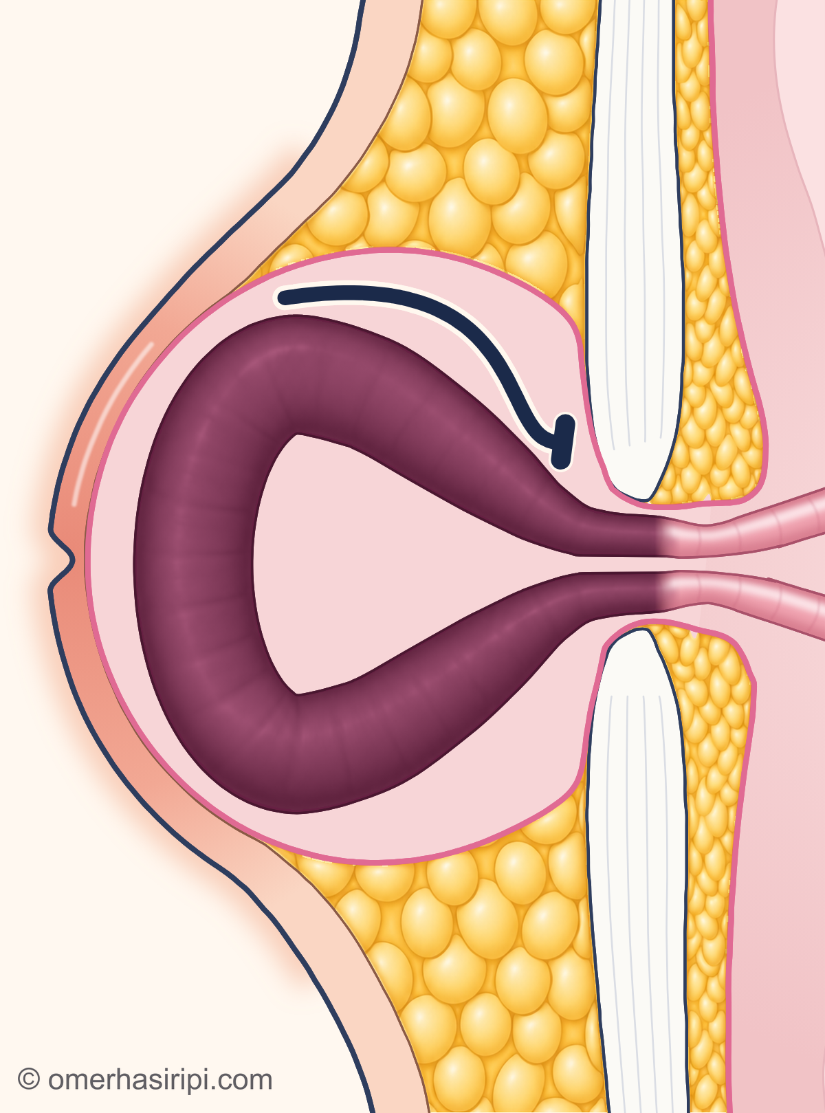
- 1 Hernia sac (saccus herniae)
- 2 Small bowel with an impaired blood supply (intestinum tenue)
- 3 Opening in the abdominal wall (anulus umbilicalis)
- 4 Cannot return inside
- 5 Firm layer where the muscle sheaths meet in the middle (linea alba)
If the blood supply to the trapped contents becomes impaired, this is called a strangulated hernia (strangulation) and urgent treatment is needed. In the drawing, the bowel with an impaired blood supply is shown in a dark colour; the skin over the hernia may also become red and painful to touch.[8, 12]
Illustrative drawing, not to scale. Prepared for this site.
Which department treats umbilical hernias?
Umbilical hernias are treated by the general surgery department. Your family doctor or workplace doctor can do the first examination; if they consider it necessary, they can refer you to general surgery.
How does an umbilical hernia go away, and does a support help?
In adults, surgery is the only way to repair an umbilical hernia, but surgery is not essential; a support or belt does not repair the hernia[8]. If your hernia is small, causes you no symptoms or very few symptoms, and you can do everything you want, including heavy lifting, work and sport, you may decide not to have surgery; this is called watchful waiting[8].
The European and American hernia societies also say that waiting may be recommended for a hernia that causes no symptoms: the evidence on this is limited, but waiting appears safe[2]. Not having surgery is safe in most cases; however, the hernia may grow and become more painful over time[8]. The NHS, on the other hand, usually recommends surgery for adults because the hernia is unlikely to go away on its own[9]. You and your surgeon decide together whether to have surgery or wait, after discussing the risks specific to you; as months and years pass, surgery may become a more suitable option[8]. While waiting, you do not need to restrict your activities unless the hernia causes a lot of discomfort during certain tasks[8].
How is umbilical hernia surgery performed, and how long does it take?
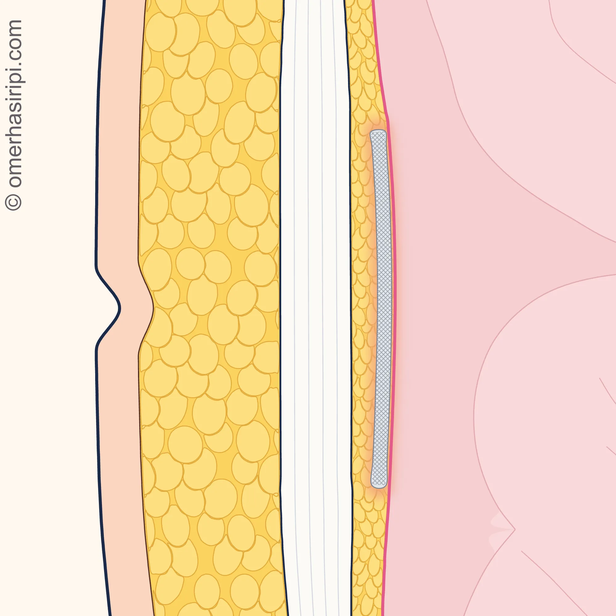
The 2020 guideline from the European and American hernia societies recommends repairing umbilical hernias that cause symptoms[1]. Using a patch (mesh) is recommended to reduce the chance of the hernia coming back; for openings smaller than 1 cm, repair with stitches alone may also be considered, with the decision made together with you[1, 2]. In open surgery, mesh does not increase wound infection[2]; repair with stitches alone, however, has a higher chance of recurrence even in small hernias[8]. The mesh can become infected and sometimes needs to be removed; however, this is very rare[8].
Most umbilical hernias can be repaired with open surgery; the mesh is preferably placed flat immediately in front of the lining of the abdomen (peritoneum)[1, 2]. Keyhole surgery (laparoscopy) may be considered if the hernia is large or your risk of wound problems is high[1]; this method reduces wound problems, but the chance of bowel injury during surgery is slightly higher[8]. The operation is usually performed under a general anaesthetic[9]; a local anaesthetic can also be used safely, and it is recommended that you, your surgeon and your anaesthetist decide together on the type of anaesthetic[3]. The operation takes 30–60 minutes[9]. For small hernias, you can usually go home the same day; for large hernias, you may need to stay in hospital for one night or several days[8].
Enlarge
Swipe the drawing right or left to see the details. You can use the arrow keys on your keyboard.
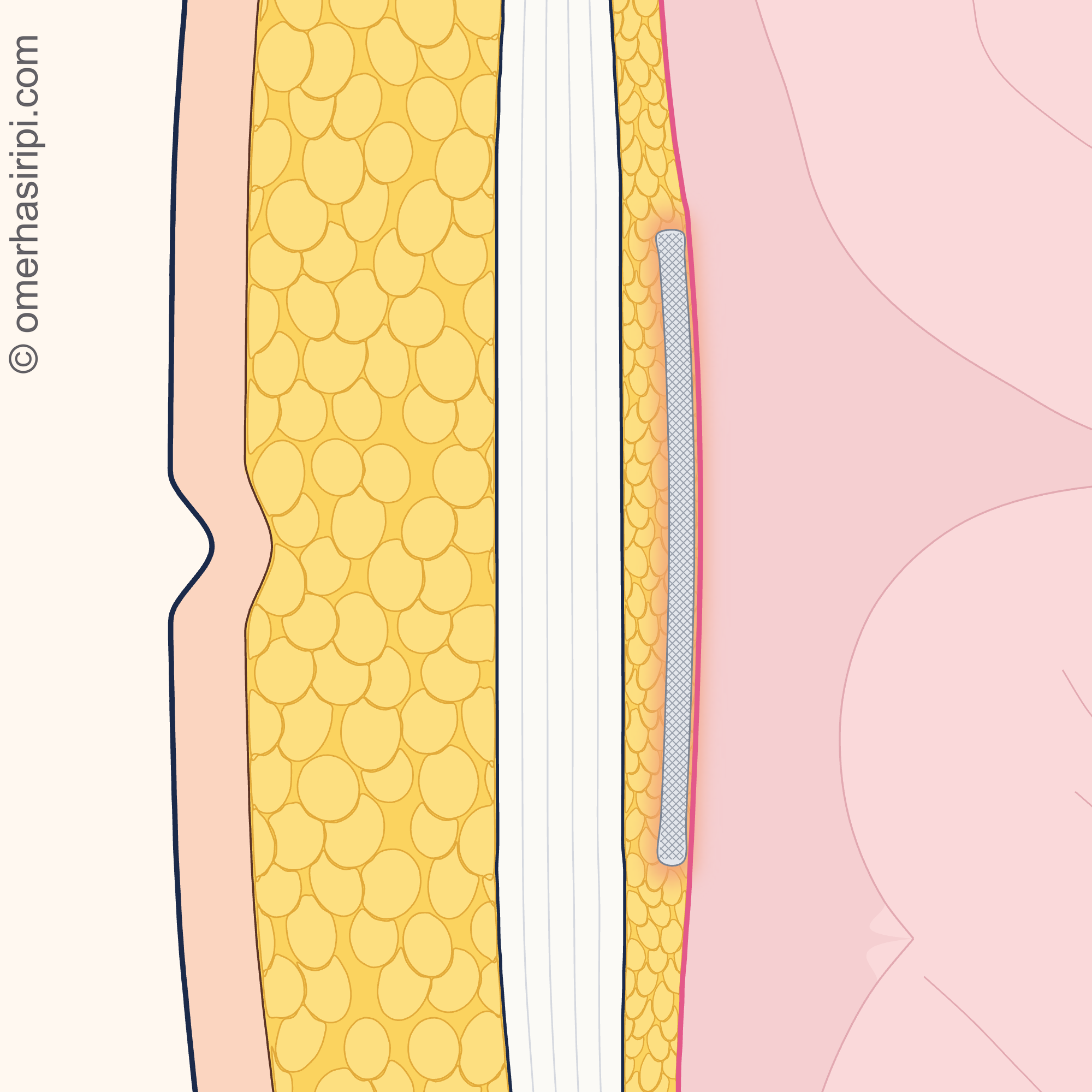
- 1 Flat patch (mesh)
- 2 Firm layer where the muscle sheaths meet in the middle
- 3 Abdominal lining (peritoneum)
- 4 Fat tissue beneath the skin
- 5 Belly button
Description of the drawing
In planned open surgery, the mesh is preferably placed flat immediately in front of the lining of the abdomen (peritoneum), behind the firm layer where the muscle sheaths meet in the middle; the mesh extends both above and below the opening. The drawing is a side section at the level of the belly button after repair: the skin is on the left and the inside of the abdomen is on the right.
Results may vary from person to person with any surgical or interventional procedure. You are advised to discuss the procedure in detail with your doctor beforehand.
Illustrative drawing, not to scale. Prepared for this site.
The rate of complications in umbilical hernia surgery is expected to be low[1]. Bleeding and a collection of fluid under the skin (seroma) are common, but almost always resolve on their own; wound infection is also common[8]. More serious complications, such as a blood clot in the leg, a blood clot travelling to the lung or bowel injury, are rare[8]. Long-lasting pain may also occur, and the hernia may come back[9]. Every operation leaves a scar, whatever the method[8].
Before surgery
Smoking and excess weight increase the risk of problems after surgery, such as wound infection and the hernia coming back[8]; however, data specific to umbilical hernias on this are limited[3]. If you smoke, you are advised to stop 4–6 weeks before surgery[3]; the European Hernia Society’s patient leaflet says at least 6 weeks[8]. If you are overweight, try to lose weight; decide with your surgeon how much to lose and over what period[8]. If you take a blood-thinning medicine, tell your surgeon; some need to be stopped temporarily days before surgery or replaced with another medicine[8]. Do not stop your medicine on your own; ask the doctor who prescribed it what to do. If you are pregnant or are considering becoming pregnant in the future, it is recommended that planned surgery be postponed until after childbirth if possible, preferably until after your last pregnancy[4].
What should you be mindful of after umbilical hernia surgery?
The general rule is to do what you want after surgery, as far as you feel comfortable[8]. Go for walks from the first day; it is recommended that someone is with you for the first few days[8]. You can usually return to desk work in 1–2 weeks and to heavy work within 2–4 weeks[8]; the NHS says that full recovery can take 4–6 weeks and that you may need to take up to 6 weeks off work if your job involves heavy lifting[9]. Your surgeon will tell you what timeframe is right for you[8].
You can drive when you can get in and out of a car, press the brake firmly without too much discomfort, and painkillers are not affecting your concentration[8]. Do not swim until your skin wounds have healed[8]. Eating fibre and drinking plenty of fluids help prevent constipation[8]. After abdominal surgery, it is common to be advised not to lift more than 5 kg for 4–6 weeks, but there is no evidence to support this[5]. In the long term, you can do what you want: there is no evidence that strenuous activities increase the chance of the hernia coming back[8].
See a doctor the same day if you have pus, redness, warmth or heavy bleeding from your wound; a fever, repeated vomiting, new or worsening shortness of breath, throbbing or cramp-like pain in your leg, or increasing abdominal pain or swelling; if you have been unable to pass stool for 3 days; or if you spit out a small amount of blood[11, 12]. Call 112 if you have severe difficulty breathing, pain in your chest or upper back, a very fast, very slow or irregular heartbeat, spit out more blood, or if your abdominal pain starts suddenly or is very severe[9, 11, 13].
Sources
15
- Henriksen NA, Montgomery A, Kaufmann R, Berrevoet F, East B, Fischer J, et al. Guidelines for treatment of umbilical and epigastric hernias from the European Hernia Society and Americas Hernia Society. Br J Surg. 2020;107(3):171–190. (The full text could not be accessed; information was taken from the abstract.) doi: 10.1002/bjs.11489. PubMed: 31916607.
- Henriksen NA, Nazari T, Simons MP, Hope W, Montgomery A. Guidelines for Treatment of Umbilical and Epigastric Hernias From the European and Americas Hernia Societies–A Web-Based Survey on Surgeons' Opinion. J Abdom Wall Surg. 2022;1:10260. (The texts of the guideline recommendations submitted for voting in 2019 are in the article’s supplementary file.) doi: 10.3389/jaws.2022.10260. PubMed: 38314164.
- European Hernia Society (EHS). Recomandări privind tratamentul herniilor ombilicale și epigastrice conform European Hernia Society and American Hernia Society Guideline (2020). Romanian translation and adaptation of the guideline in Source 1 (translated by Ioana Florea). Text: europeanherniasociety.eu.
- Henriksen NA, Kaufmann R, Simons MP, Berrevoet F, East B, Fischer J, et al. EHS and AHS guidelines for treatment of primary ventral hernias in rare locations or special circumstances. BJS Open. 2020;4(2):342–353. doi: 10.1002/bjs5.50252. PubMed: 32207571.
- Hernández-Granados P, Henriksen NA, Berrevoet F, Cuccurullo D, López-Cano M, Nienhuijs S, et al. European Hernia Society guidelines on management of rectus diastasis. Br J Surg. 2021;108(10):1189–1191. (The full text of the guideline is in the article’s supplementary file.) doi: 10.1093/bjs/znab128. PubMed: 34595502.
- Birindelli A, Sartelli M, Di Saverio S, Coccolini F, Ansaloni L, van Ramshorst GH, et al. 2017 update of the WSES guidelines for emergency repair of complicated abdominal wall hernias. World J Emerg Surg. 2017;12:37. doi: 10.1186/s13017-017-0149-y. PubMed: 28804507.
- Song DS, Yoo JJ, Song JE, Kang SH, Shin SK, Han SK, et al. KASL clinical practice guidelines for liver cirrhosis: Ascites and related complications 2026. Clin Mol Hepatol. 2026;32(3):961–1028. doi: 10.3350/cmh.2026.0281. PubMed: 42050795.
- East B, Henriksen N, Dames N, Pawlak M, East MJ, Montgomery A, et al. European Hernia Society (EHS). Umbilical and epigastric hernia: information for patients (patient leaflet based on the EHS and AHS guidelines). February 2020. A Turkish translation by the Turkish Hernia Society is also available. Text: europeanherniasociety.eu.
- NHS (National Health Service of England). Umbilical hernia (umbilical hernia): patient information page. Last reviewed: 24 November 2025. Text: www.nhs.uk.
- NHS (National Health Service of England). Hernia (hernia): patient information page for all types of hernia. Last reviewed: 19 May 2026. Text: www.nhs.uk.
- NHS (National Health Service of England). Inguinal hernia repair (groin hernia surgery): patient information page. Last reviewed: 28 February 2025. Text: www.nhs.uk.
- American College of Surgeons (ACS). Ventral Hernia Repair (abdominal wall hernia surgery): patient information page that also covers umbilical hernias. Last updated: 2024. Text: www.facs.org.
- NHS (National Health Service of England). Stomach ache (abdominal pain): patient information page. Last reviewed: 26 May 2023 (the next review is overdue). Text: www.nhs.uk.
- OpenStax. Anatomy and Physiology 2e. 2022. Section 11.4: Axial Muscles of the Abdominal Wall, and Thorax; full text accessed 3 October 2026. Text: openstax.org.
- OpenStax. Anatomy and Physiology 2e. 2022. Section 23.1: Overview of the Digestive System; full text accessed 28 October 2026. Text: openstax.org.
Medical review of the Turkish original: Op. Dr. Ömer Seyfullah Hasıripi, . This page is a translation checked by AI reviewers; the Turkish version prevails.
This page is for general information; it does not replace an examination. See a doctor about your symptoms.