If you have a gastroscopy appointment: Follow the preparation instructions given to you by the unit performing the procedure; if you have no instructions, ask the unit[1]. If you are pregnant or take regular medicines (such as blood thinners or diabetes medicine), tell the unit as soon as you receive the instructions[1, 2, 3]. Do not stop your medicines without consulting the unit or the doctor who prescribed them[3, 4].
What is gastroscopy (stomach endoscopy)? Is it the same as endoscopy?
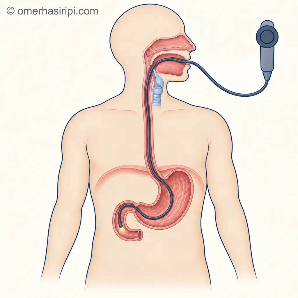
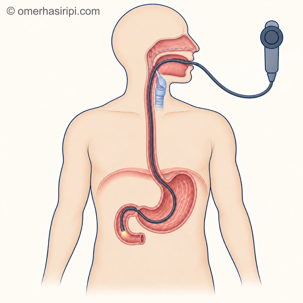
Gastroscopy (stomach endoscopy) is an examination in which a thin, flexible tube with a small camera at its tip is used to look inside the oesophagus, stomach and duodenum (the first part of the small intestine)[1, 3]. The other medical name for this examination is oesophagogastroduodenoscopy[1, 2, 3]. In everyday speech, “endoscopy” usually means this examination; however, endoscopy is also the general name for similar examinations, such as colonoscopy, which looks inside the large bowel.
The camera can show problems such as inflammation, sores (ulcers), polyps and tumours[3]. If needed, a small tissue sample (biopsy) may be taken, a polyp removed, bleeding stopped or a narrowed area widened during the same procedure[1, 3].
Why is gastroscopy done?
It is done to find the cause of symptoms such as indigestion, difficulty swallowing, abdominal pain, black and sticky stools or vomiting blood, to treat certain problems in the digestive system and to investigate stomach or oesophageal cancer[1]. If you have sudden and obvious gastrointestinal bleeding (for example, vomiting blood) along with indigestion, call 112 or go to the nearest emergency department[15].
If you have difficulty swallowing, or if you are aged 55 or over and are losing weight along with indigestion, reflux or upper abdominal pain, see a doctor without delay: British guidelines call for prompt investigation for oesophageal and stomach cancer in these situations[5, 6]. If there is oesophageal or stomach cancer in your family, the age threshold is lower: the British Society of Gastroenterology recommends prompt gastroscopy for people with indigestion in this group who are over 40[6]. The North American guideline uses age 60 as the threshold for gastroscopy in indigestion and states that it may be done earlier in people at high risk of cancer[7].
Outside these situations, most people with indigestion do not need a gastroscopy straight away[6]. Guidelines recommend that these people are first tested for the stomach bacterium (Helicobacter pylori) with a breath or stool test and treated if it is present; if the test is negative, they recommend trying a medicine that reduces stomach acid[6, 7]. About 8 in 10 people who have a gastroscopy for indigestion are diagnosed with functional dyspepsia, meaning no tissue disease is found to explain their symptoms[6]; the likelihood of finding something serious, such as cancer, is low[6].
If a gastroscopy recommended by your doctor is not done, the diagnosis of conditions such as an ulcer or stomach tumour may be delayed[3].
How many hours should you fast before gastroscopy, and can you drink water?
Follow the fasting time given to you by the unit[1]. According to the European endoscopy society, solid food is stopped at least 6 hours and clear liquids at least 2 hours before the procedure[8]. Your unit may ask for a longer period: the Turkish Society of Gastroenterology’s consent form requires at least 8 hours of fasting[3]; fasting for 8 hours or longer may also be needed after a heavy, fatty meal[9].
Clear liquids such as water, tea, coffee (up to one fifth milk may be added) and fruit juice without pulp can be drunk until 2 hours before the procedure; for milk and milky drinks, the interval is 6 hours[9]. As sources differ on water, ask the unit whether you can drink it[1, 3, 9].
If gastroscopy is to be done to confirm coeliac disease, continue eating foods containing gluten at more than one meal a day for at least 6 weeks before the procedure, so that the result does not mistakenly appear normal[2].
Can you take blood pressure medicine and other medicines?
Bring a list of all the medicines you take and follow the unit’s instructions about medicines[1]. Ask the unit how to take your regular medicines, such as blood pressure medicine, on the morning of the procedure, whether you can smoke and whether any tests are needed.
What to do with blood thinners depends on the medicine and the procedure to be performed; the joint guideline from the British and European societies and the Turkish consent form also give different recommendations[3, 4]. Stopping the medicine carries a risk of clotting problems such as stroke, while continuing it carries a risk of bleeding[4]. The unit or the doctor who prescribed the medicine will decide what you should do after discussing it with you; if a change is needed, you should be told in writing and verbally what to do and when[2, 3, 4].
If you take medicine for diabetes or weight loss (including injections), tell the unit about this too[1, 10]. The role of GLP-1 medicines before surgery and endoscopy. Also tell them if you smoke or drink alcohol, snore or stop breathing in your sleep (sleep apnoea), have an allergy to a medicine, take painkillers or have previously had problems with a sedative or general anaesthetic; these may affect the sedative[3, 11].
How is gastroscopy done, and how long does it take?
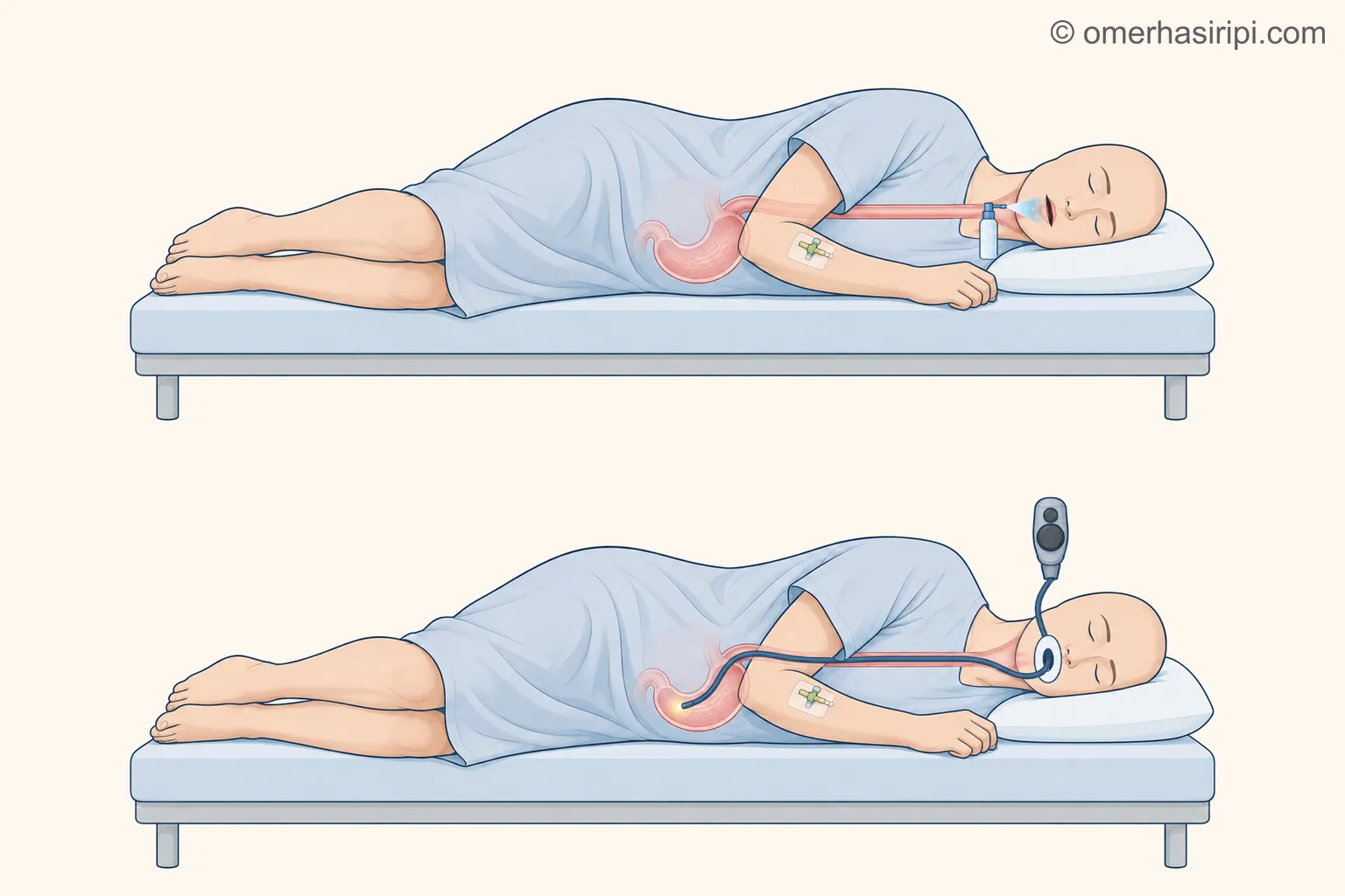
To make the procedure easier, you may be offered a spray to numb your throat, a sedative given through a thin tube in your arm to help you relax, or both[1, 2]. It can also be done with the spray alone, without a sedative[3, 10]; you and your doctor decide together which to choose[10, 11]. According to the consent form, the sedative makes you drowsy: you do not lose consciousness completely, but most of the time you will not remember the procedure in detail afterwards[3].
If you have dentures or glasses, these are removed, and you lie on your left side[3]. The tube enters through your mouth or nose and passes through your throat to your stomach[1]. If it is to enter through your mouth, a mouthpiece is placed to keep your mouth open[3]. You may feel as if you are going to vomit until the tube reaches your stomach; the sedative helps prevent this[1]. Because the stomach is inflated with air, you may feel bloated and burp a few times[1]. You can breathe comfortably throughout the procedure[3]. You will not feel anything when a tissue sample is taken[1]. There may be some discomfort; you can ask for the procedure to be stopped at any time[1, 11].
Enlarge
Swipe the drawing right or left to see the details. You can use the arrow keys on your keyboard.
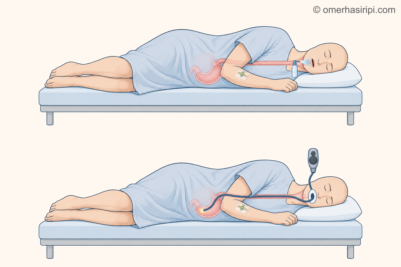
- 1 Throat-numbing spray
- 2 Thin tube in the arm
- 3 Thin, flexible tube
- 4 Mouthpiece
- 5 Oesophagus
- 6 Stomach
- 7 Camera at the tip of the tube
- 8 Endoscope handle
Description of the drawing
You lie on your left side. The throat-numbing spray is shown at the top; the thin tube in your arm is where the sedative is given. Below, the thin, flexible tube enters through the mouthpiece and passes through your throat into your oesophagus and stomach; the camera at its tip shows the inside.
Results may vary from person to person with any surgical or interventional procedure. You are advised to discuss the procedure in detail with your doctor beforehand.
Illustrative drawing, not to scale. Prepared for this site.
The doctor or nurse may explain the result before you leave the unit; if a sample was taken, you will wait separately for its result[1]. If this result does not arrive within the time you were told, call the unit[16].
How many minutes does a gastroscopy take?
The reported length of the procedure varies by source: the Turkish consent form says an average of 5–10 minutes[3], while the patient information page from England says 15–45 minutes[1]. The European endoscopy society requires the tube to remain inside for at least 7 minutes during a diagnostic examination[8]. The total time you spend at the unit is longer: according to the patient page from England, it can take about 4 hours from arrival to returning home[1].
When can you eat after gastroscopy, and what should you consider?
If your throat was sprayed, do not eat or drink anything until the numbness wears off, about 1 hour[1, 3]. If you only had a sedative, wait 30–45 minutes; unless the unit tells you otherwise, you can eat and drink once your swallowing returns to normal[16]. If you had a sedative, someone must take you home and stay with you for 24 hours[1, 3, 10]. During these 24 hours, do not drink alcohol, drive or operate machinery, or sign important documents[1]. Do not take sleeping tablets either[16]; if you take them regularly, tell the unit before the procedure and follow its instructions[1].
You may have some pain in your abdomen and throat; this is normal[1]. Abdominal pain often goes away within a few hours[1]; a sore throat usually eases within 2 days[16]. Bloating decreases as you pass wind[3]. A very small amount of blood may come from your mouth because your throat is irritated or because samples were taken[3]. If the bloating or pain in your abdomen lasts longer than 24 hours, or if you vomit, call the unit where the procedure was performed immediately; if you cannot reach it, go to the emergency department[3, 16]. Ask for written information and a telephone number before leaving the unit[10].
What are the risks of gastroscopy?
The risk of gastroscopy is low, but it is not zero[3, 12]. In large studies, an adverse event was reported at rates ranging from 1 in 200 to 1 in 10,000 procedures, and death at rates ranging from none to 1 in 2,000 procedures[12]. Up to 60% of these events are heart and breathing problems related to sedatives and painkillers; this risk is higher in people with pre-existing heart or lung disease and in older people[12]. Perforation has been reported at rates between 1 in 2,500 and 1 in 11,000 procedures; significant bleeding is rare in gastroscopy performed only to examine[12]. The risk of bleeding and perforation may increase with procedures such as polyp removal, widening a narrowing or stopping bleeding[3]; bleeding may also occur 1–2 weeks later[4]. Very rarely, teeth may be damaged[3, 13]; if you have a loose tooth, say so beforehand[13]. If the tube enters through the nose, the nose may bleed; say so beforehand if you have had heavy or frequent nosebleeds, nasal surgery or a nasal injury[13]. Gastroscopy may not show every condition: a review of studies reported that about 1 in 10 upper digestive tract cancers had been missed at gastroscopy in the 3 years before diagnosis[13]; if your symptoms persist or worsen, see a doctor again[5].
Which department deals with gastroscopy?
Gastroenterology specialists perform gastroscopy; in Turkey, general surgery specialists trained in this area can also perform it[14]. For symptoms such as indigestion, your family doctor or workplace doctor may carry out the initial assessment[6]; if they consider it necessary, they may refer you to the appropriate department.
Sources
16
- NHS (National Health Service in England). Gastroscopy: patient information page. Last reviewed: 13 August 2026. Text: www.nhs.uk.
- Beg S, Ragunath K, Wyman A, Banks M, Trudgill N, Pritchard DM, et al. Quality standards in upper gastrointestinal endoscopy: a position statement of the British Society of Gastroenterology (BSG) and Association of Upper Gastrointestinal Surgeons of Great Britain and Ireland (AUGIS). Gut. 2017;66(11):1886–1899. doi: 10.1136/gutjnl-2017-314109. PubMed: 28821598.
- Türk Gastroenteroloji Derneği. Üst gastrointestinal sistem endoskopisi (özofagogastroduodenoskopi) onam formu. The society’s sample consent form (Word file); it is not a guideline and the text is undated. Text: tgd.org.tr.
- Veitch AM, Radaelli F, Alikhan R, Dumonceau JM, Eaton D, Jerrome J, et al. Endoscopy in patients on antiplatelet or anticoagulant therapy: British Society of Gastroenterology (BSG) and European Society of Gastrointestinal Endoscopy (ESGE) guideline update. Gut. 2021;70(9):1611–1628. doi: 10.1136/gutjnl-2021-325184. PubMed: 34362780.
- National Institute for Health and Care Excellence (NICE). Suspected cancer: recognition and referral (NG12). Published: 23 June 2015; last updated: 15 April 2026. Text: www.nice.org.uk.
- Black CJ, Paine PA, Agrawal A, Aziz I, Eugenicos MP, Houghton LA, et al. British Society of Gastroenterology guidelines on the management of functional dyspepsia. Gut. 2022;71(9):1697–1723. doi: 10.1136/gutjnl-2022-327737. PubMed: 35798375.
- Moayyedi P, Lacy BE, Andrews CN, Enns RA, Howden CW, Vakil N. ACG and CAG Clinical Guideline: Management of Dyspepsia. Am J Gastroenterol. 2017;112(7):988–1013. (Recommendations taken from the article's abstract.) doi: 10.1038/ajg.2017.154. PubMed: 28631728.
- Areia M, Esposito G, Leclercq P, Romańczyk M, Zessner-Spitzenberg J, Delgado Guillena PG, et al. Performance measures for upper gastrointestinal endoscopy: a European Society of Gastrointestinal Endoscopy (ESGE) Quality Improvement Initiative – Update 2025. Endoscopy. 2025;57(11):1268–1297. (Recommendations taken from the article's abstract.) doi: 10.1055/a-2674-4912. PubMed: 40934951.
- Rüggeberg A, El-Boghdadly K, Bilotta F, Vaz MD, Camilleri Podesta AM, Jammer I, et al. Peri-operative fasting in adults: an international, multidisciplinary consensus statement. Anaesthesia. 2026;81(6):840–851. (International consensus statement in the field of anaesthesia.) doi: 10.1111/anae.70130. PubMed: 41657234.
- Triantafyllou K, Tziatzios G, Tham TC, Beilenhoff U, Lorenzo-Zúñiga V, Pouw RE, et al. Sedation for gastrointestinal endoscopy: European Society of Gastrointestinal Endoscopy (ESGE) and European Society of Gastroenterology and Endoscopy Nurses and Associates (ESGENA) Guideline. Endoscopy. 2026;58(10):1142–1179. (Recommendations taken from the article's abstract.) doi: 10.1055/a-2898-6540. PubMed: 42480549.
- ASGE Standards of Practice Committee; Early DS, Lightdale JR, Vargo JJ 2nd, Acosta RD, Chandrasekhara V, Chathadi KV, et al. Guidelines for sedation and anesthesia in GI endoscopy. Gastrointest Endosc. 2018;87(2):327–337. doi: 10.1016/j.gie.2017.07.018. PubMed: 29306520.
- ASGE Standards of Practice Committee; Ben-Menachem T, Decker GA, Early DS, Evans J, Fanelli RD, Fisher DA, et al. Adverse events of upper GI endoscopy. Gastrointest Endosc. 2012;76(4):707–718. doi: 10.1016/j.gie.2012.03.252. PubMed: 22985638.
- Waddingham W, Kamran U, Kumar B, Trudgill NJ, Tsiamoulos ZP, Banks M. Complications of diagnostic upper Gastrointestinal endoscopy: common and rare – recognition, assessment and management. BMJ Open Gastroenterol. 2022;9(1):e000688. (Review; not a guideline.) doi: 10.1136/bmjgast-2021-000688. PubMed: 36572454.
- Türk Cerrahi Derneği. Endoskopi hakkında bilgilendirme (the association’s announcement to its members; undated). Text: www.turkcer.org.tr.
- National Institute for Health and Care Excellence (NICE). Gastro-oesophageal reflux disease and dyspepsia in adults: investigation and management (CG184). Published: 3 September 2014; last updated: 18 October 2019. Text: www.nice.org.uk.
- Guy's and St Thomas' NHS Foundation Trust (London). After a gastroscopy: patient information page, 4846/VER4. Last reviewed: April 2026. Accessed: 29 September 2026. Text: www.guysandstthomas.nhs.uk.
Medical review of the Turkish original: Op. Dr. Ömer Seyfullah Hasıripi, . This page is a translation checked by AI reviewers; the Turkish version prevails.
This page is for general information; it does not replace an examination. See a doctor about your symptoms.