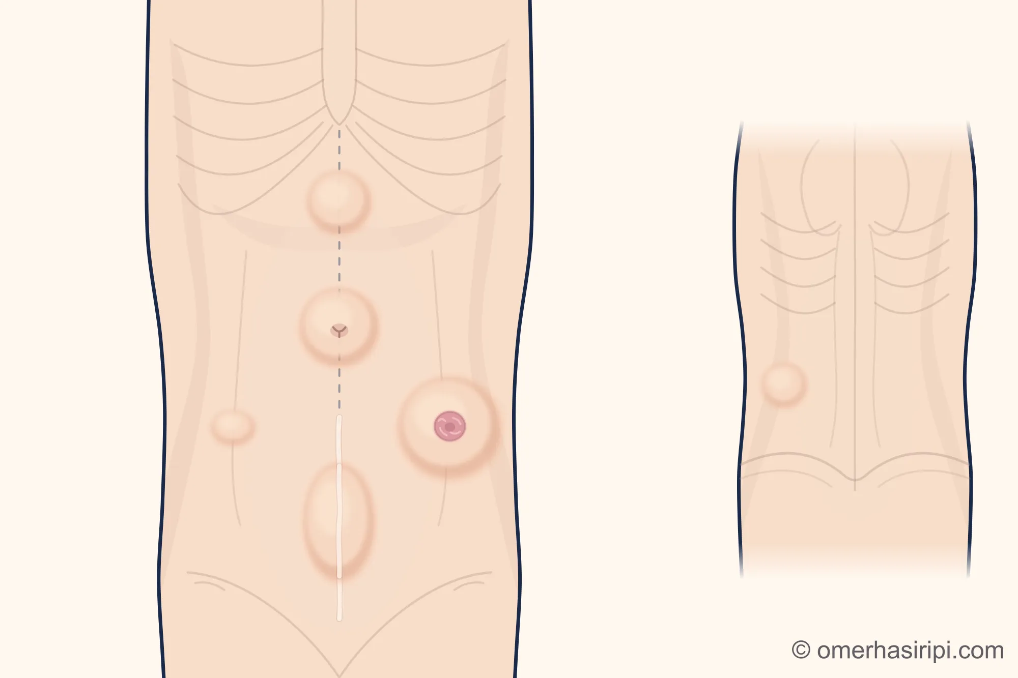
What is an abdominal hernia and what types are there?
A hernia forms when fat tissue or organs inside the abdomen are pushed out through an opening in the abdominal wall[5, 9]. Their names vary according to their location[5, 9, 10]:
- An umbilical hernia forms at the navel; details are explained on its own page[5, 11].
- An upper abdominal hernia (epigastric hernia) is found on the midline between the navel and the breastbone[4, 9].
- An incisional hernia (surgical-site hernia) develops beneath an old abdominal surgery scar[5, 9].
- A Spigelian hernia develops on the side of the abdomen; a lumbar hernia develops in the back and side part of the abdominal wall at the waist[5, 7, 8].
- A parastomal hernia is around an opening made in the abdominal wall for the bowel or urinary tract[10].
Separation of the abdominal muscles at the midline (rectus diastasis) is not a true hernia[6].
What causes an incisional hernia and when does it appear?
An incisional hernia is a common problem after abdominal surgery[2]. The European Hernia Society lists a high body mass index (excess weight for your height), smoking, diabetes and surgical wound infection among the risk factors[1]. An incision made along the midline carries a higher risk than one made away from the midline; the way the incision is closed also affects the risk[1]. The American College of Surgeons’ patient information gives the rate of incisional hernia after abdominal surgery as 5–17%; most appear within the first five years[9]. This range does not mean the likelihood is the same for every operation or every person[1, 9].
What are the symptoms of an abdominal hernia and how is it diagnosed?
The most noticeable sign is a bulge; it may become more prominent when coughing or straining and may be accompanied by pain or a feeling of pressure[9]. A Spigelian hernia, however, can sometimes cause symptoms without a bulge that can be felt[7]. With a hernia above the navel, the bulge may be more visible when standing and become smaller when lying down[5]. If an incisional hernia is suspected and an examination does not make the diagnosis clear, imaging is recommended; computed tomography is the most sensitive test[1]. If surgery is being considered, a CT or MRI scan may be used to plan the repair of an incisional hernia[1]. A hernia above the navel is usually diagnosed by examination; if there is doubt, an ultrasound or CT scan may be needed[4, 5].
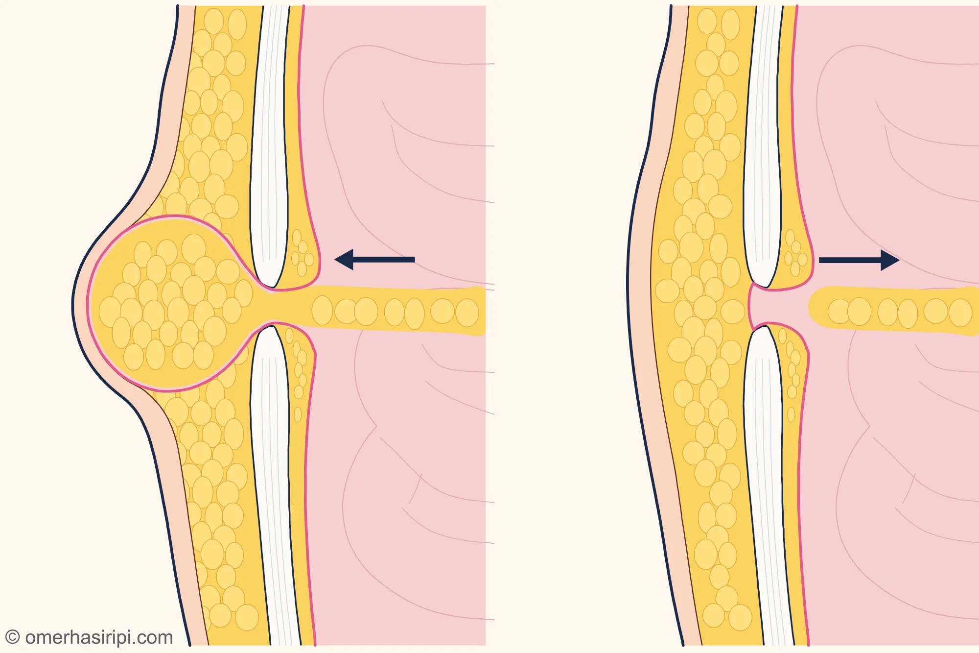
Enlarge
Swipe the drawing right or left to see the details. You can use the arrow keys on your keyboard.
- 1 When standing
- 2 When lying down
- 3 Opening in the abdominal wall
- 4 Fatty tissue pushed outwards
- 5 Firm layer where the muscle sheaths meet in the middle
- 6 Abdominal lining (peritoneum)
- 7 Fat tissue sliding back
Description of the drawing
Side cross-section of the abdominal wall: the skin is on the left and the inside of the abdomen is on the right. When standing, tissue inside the abdomen is pushed out through the opening and the bulge becomes more prominent; when lying down, it may slide back, and the bulge becomes smaller or disappears. Even if the bulge disappears, the opening has not been repaired; surgery is the way to correct the hernia.[5, 9]
Illustrative drawing, not to scale. Prepared for this site.
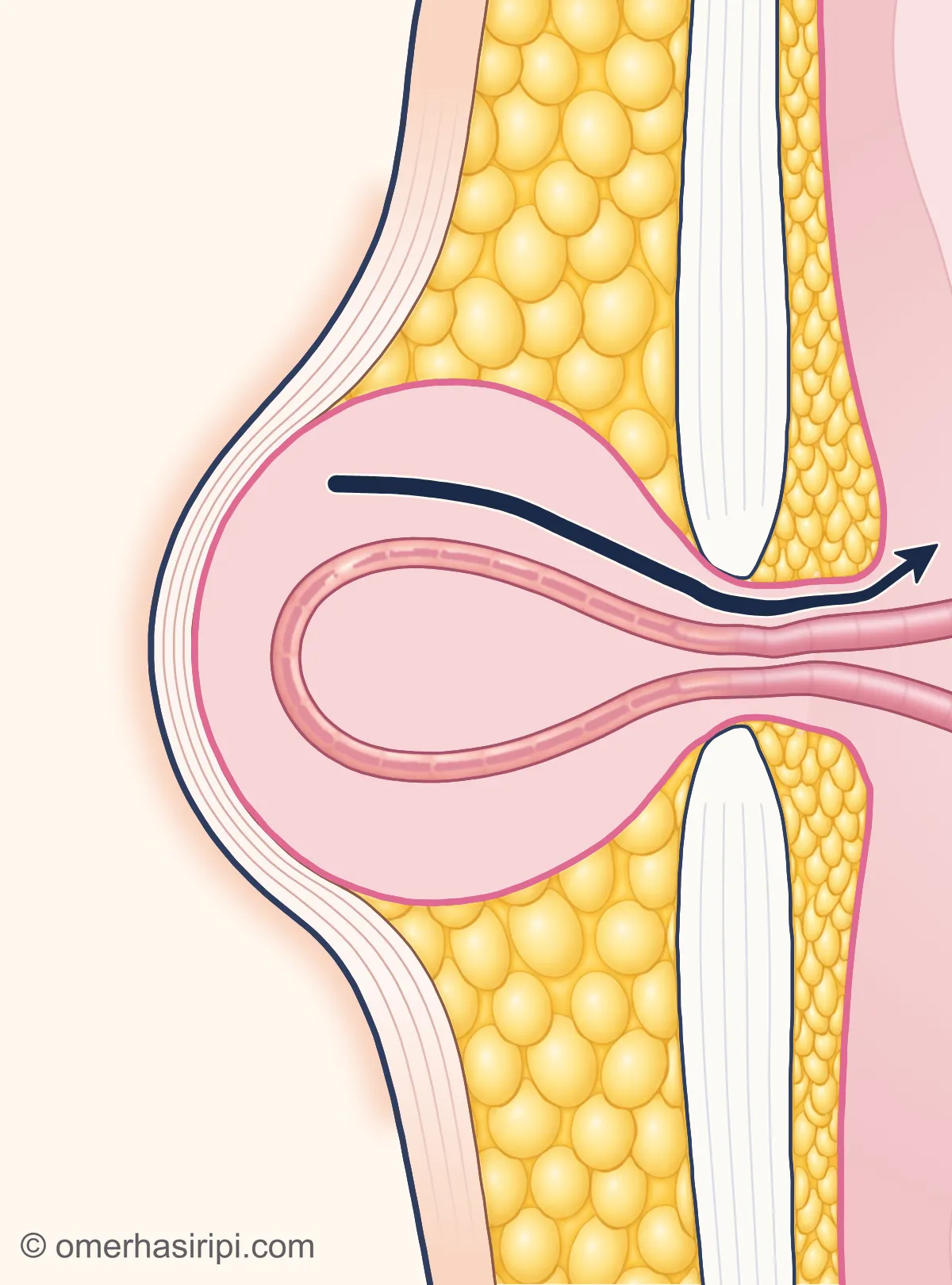
Enlarge
Swipe the drawing right or left to see the details. You can use the arrow keys on your keyboard.
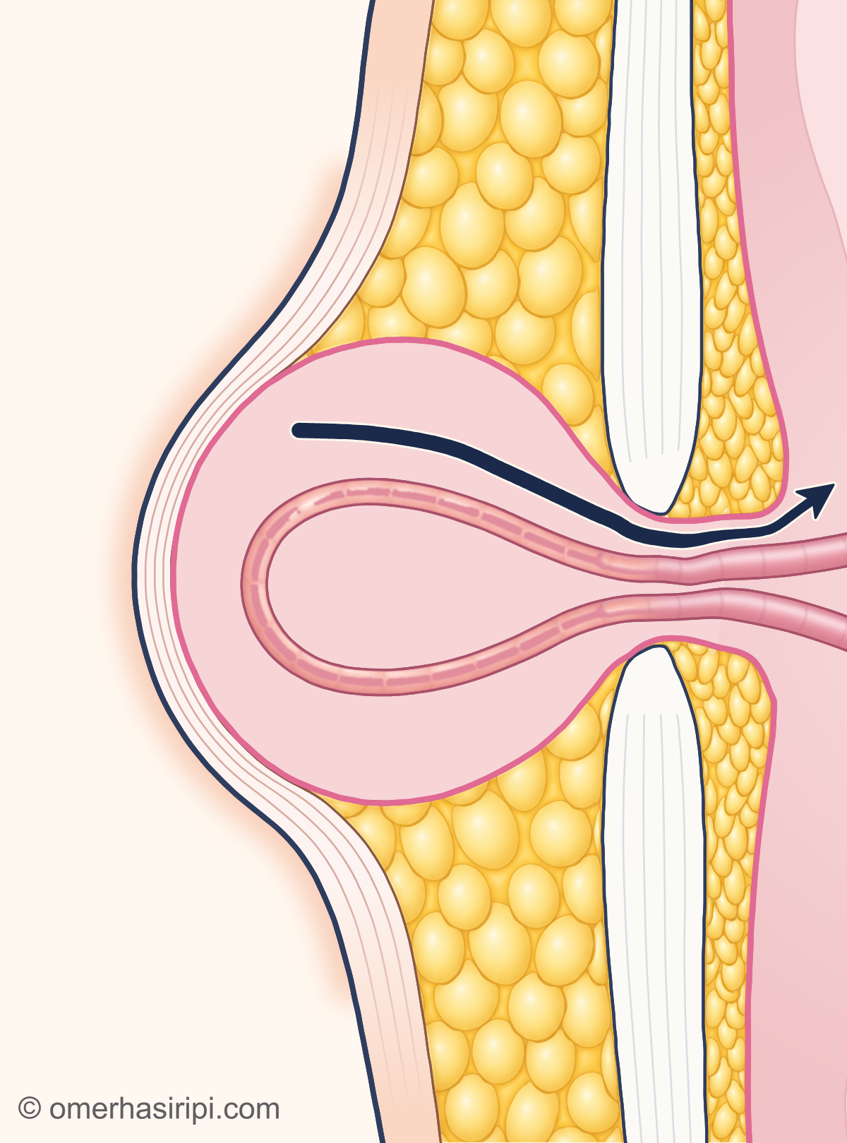
- 1 Hernia sac (saccus herniae)
- 2 Small bowel pushed outwards (intestinum tenue)
- 3 Opening in the abdominal wall
- 4 Can return inside
- 5 Firm layer where the muscle sheaths meet in the middle (linea alba)
- 6 Old surgical scar
Description of the drawing
A hernia may also contain bowel. If it becomes smaller when you lie down or press on it gently with your hand, this means the hernia can go back in.[9]
Illustrative drawing, not to scale. Prepared for this site.
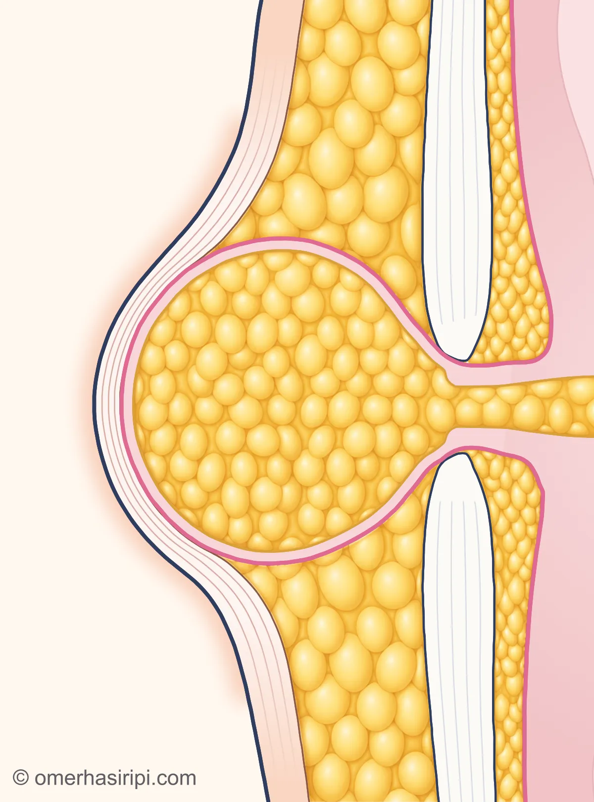
Enlarge
Swipe the drawing right or left to see the details. You can use the arrow keys on your keyboard.
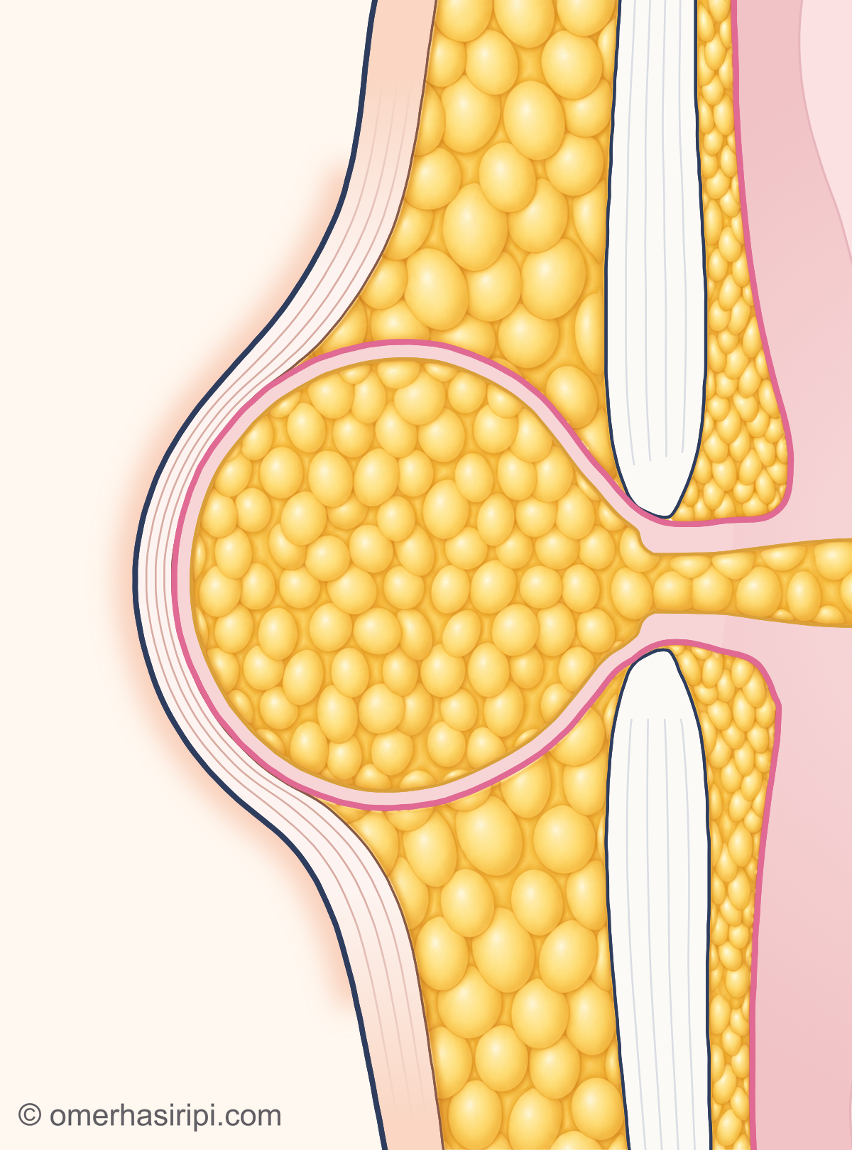
- 1 Hernia sac (saccus herniae)
- 2 Fatty apron inside the abdomen (omentum majus)
- 3 Opening in the abdominal wall
- 4 Fat tissue beneath the skin
- 5 Firm layer where the muscle sheaths meet in the middle (linea alba)
- 6 Old surgical scar
Description of the drawing
A hernia may also contain fatty tissue instead of bowel. In the drawing, this fat is a part of the fatty apron inside the abdomen (omentum) that has been pushed into the sac.[15, 16]
Illustrative drawing, not to scale. Prepared for this site.
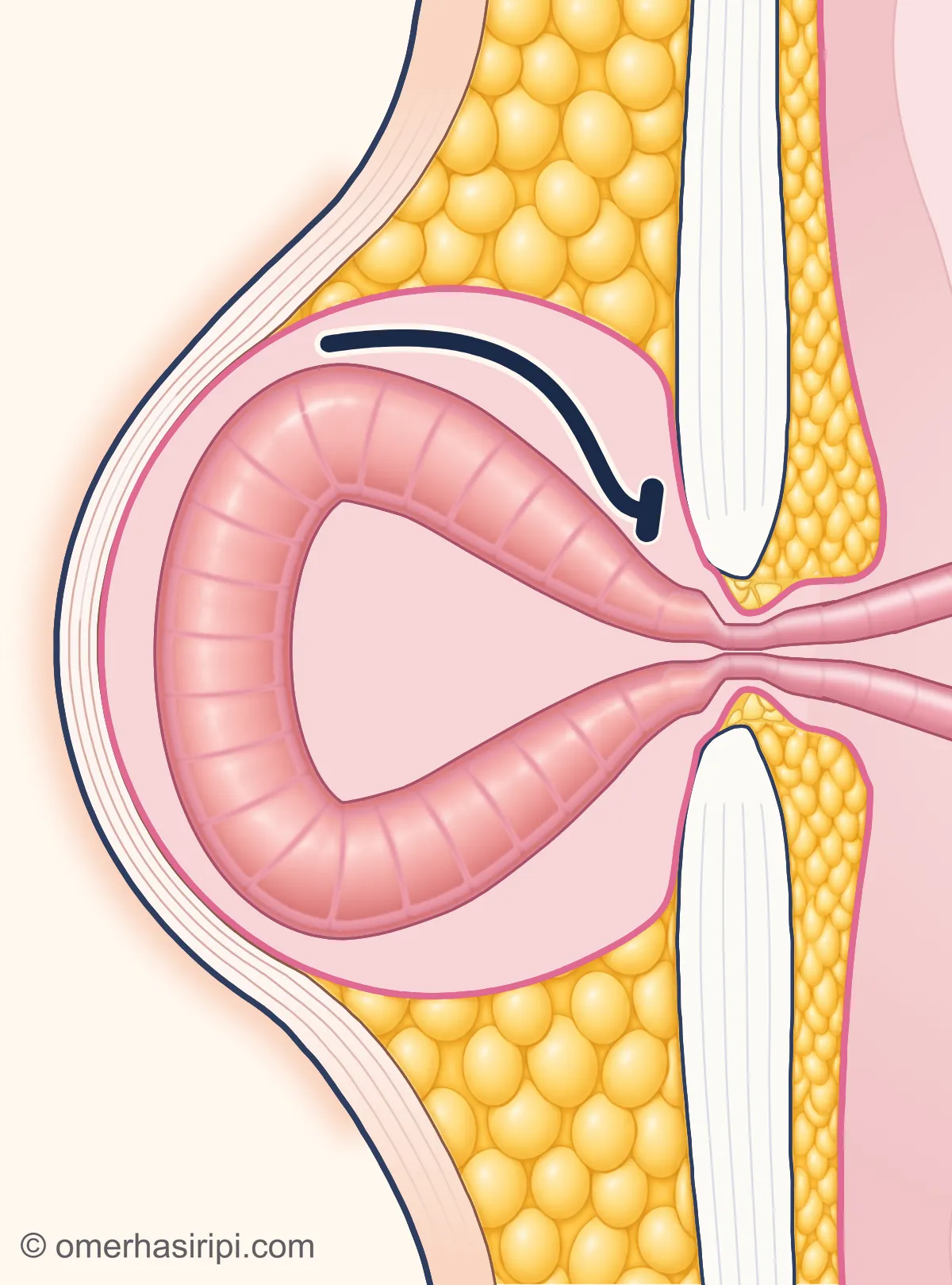
Enlarge
Swipe the drawing right or left to see the details. You can use the arrow keys on your keyboard.
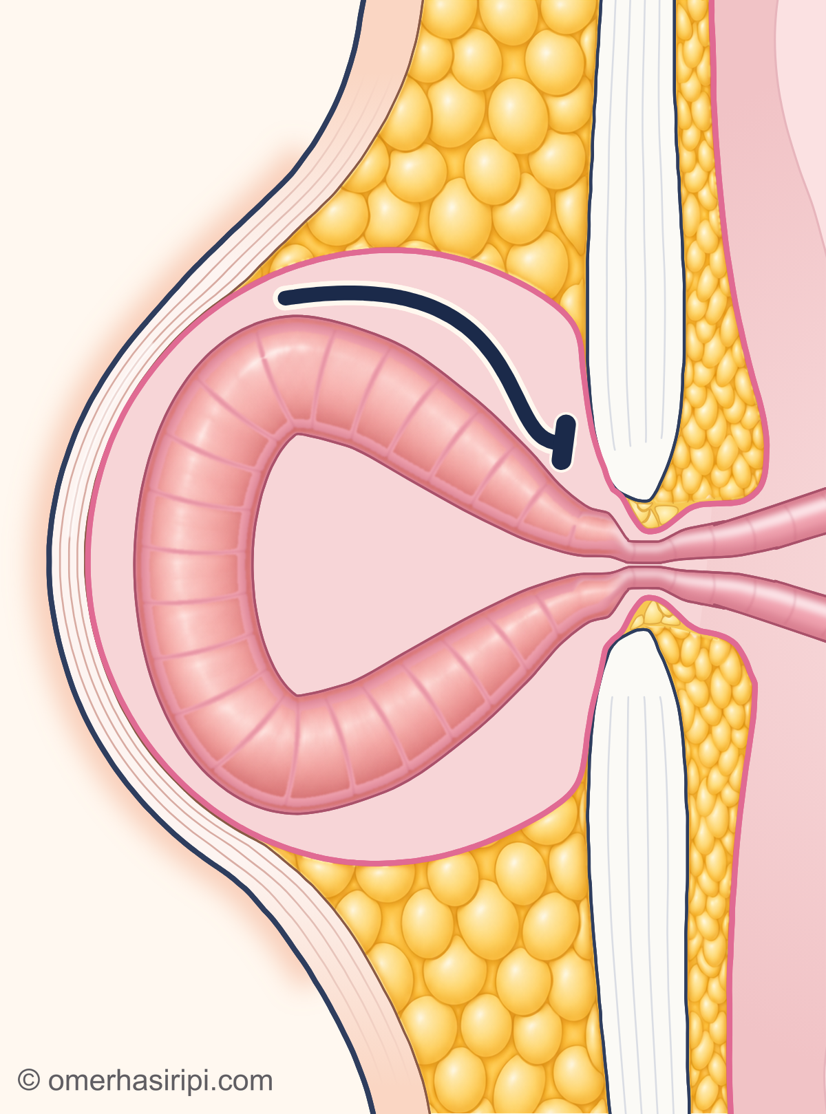
- 1 Hernia sac (saccus herniae)
- 2 Small bowel trapped in the hernia (intestinum tenue)
- 3 Opening in the abdominal wall
- 4 Cannot return inside
- 5 Firm layer where the muscle sheaths meet in the middle (linea alba)
- 6 Old surgical scar
If the contents become trapped in the sac and cannot go back into the abdomen, the hernia has become trapped (incarceration). A narrow opening or the contents sticking to the sac can cause this; bowel obstruction may also develop. Sudden, sharp abdominal pain and vomiting may indicate this; if you have these symptoms, follow the instructions in the “When is it an emergency?” section.[9, 16]
Illustrative drawing, not to scale. Prepared for this site.
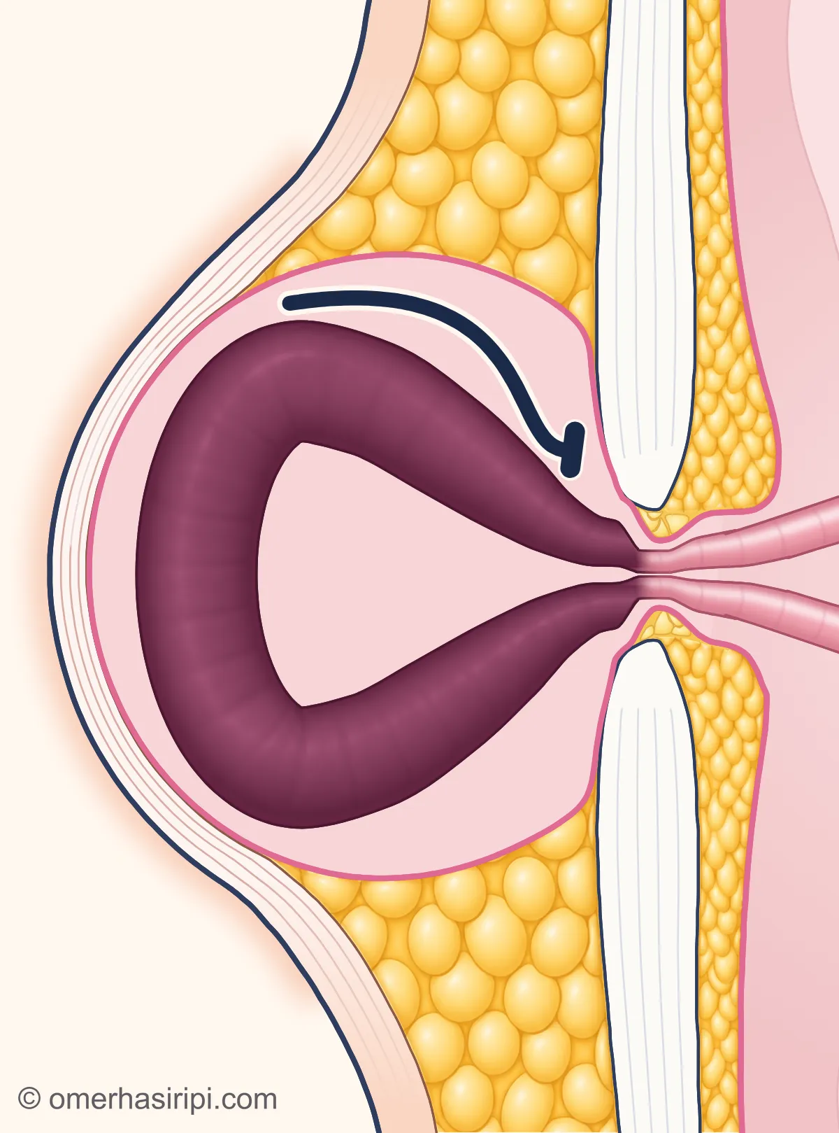
Enlarge
Swipe the drawing right or left to see the details. You can use the arrow keys on your keyboard.
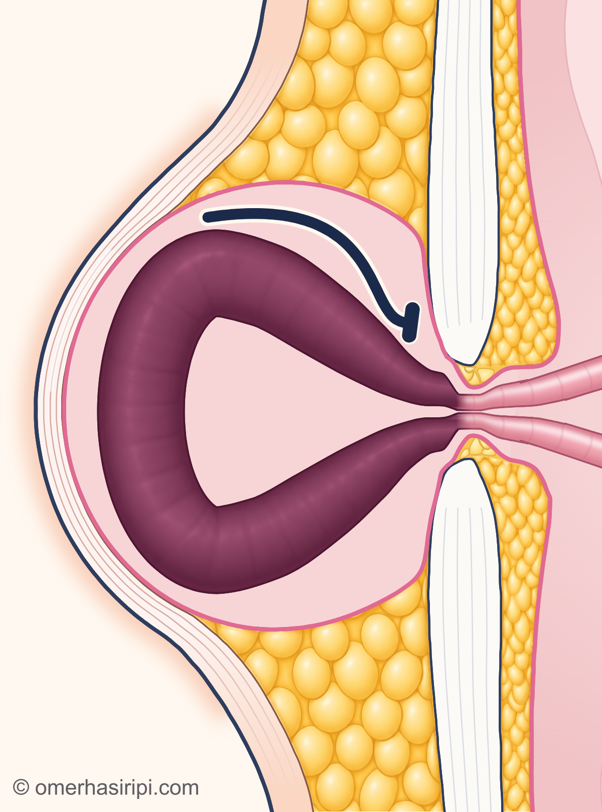
- 1 Hernia sac (saccus herniae)
- 2 Small bowel with an impaired blood supply (intestinum tenue)
- 3 Opening in the abdominal wall
- 4 Cannot return inside
- 5 Firm layer where the muscle sheaths meet in the middle (linea alba)
- 6 Old surgical scar
If the blood supply to the contents of the hernia is impaired, this is called a strangulated hernia (strangulation). If you have a hernia and experience sharp abdominal pain together with vomiting, the bowel may be trapped in the hernia and its blood supply may be impaired; this is a surgical emergency and requires immediate treatment. In the drawing, the bowel with impaired blood supply is shown in a dark colour.[9, 16]
Illustrative drawing, not to scale. Prepared for this site.
Will an abdominal hernia go away with a support belt, or is surgery needed?
In adults, a support belt may provide comfort, but it does not repair the opening; surgery is the way to correct the hernia[9]. According to the European Hernia Society, for a midline incisional hernia that can be pushed back in by hand, the risk of strangulation of the hernia (impaired blood supply to trapped tissue) or bowel obstruction is 1% in the first year and 2.5% over five years[1]. These rates have been reported only for this type of hernia[1]. For a Spigelian hernia, planned repair is recommended even if there are no symptoms because of the risk of tissue becoming trapped[7]. For a lumbar hernia that develops spontaneously, planned repair is also recommended as early as possible[8]. If you have a midline incisional hernia, your health is suitable for surgery and your symptoms affect your quality of life, discuss the benefits and risks of surgery and monitoring with your surgeon and consider repair[1]. Surgery carries risks such as the hernia coming back, infection, injury to internal organs and ongoing pain[9]. Monitoring may be considered for a hernia above the navel without symptoms; the Romanian EHS/AHS adaptation recommends repair for a hernia that causes symptoms[3, 4]. If the hernia grows or becomes painful during monitoring, it would be appropriate to seek a doctor’s advice[5].
How do you prepare for abdominal hernia surgery?
Before planned incisional hernia surgery, targeted weight loss if you are overweight, good control of diabetes and stopping smoking are recommended[1]. If you have an incisional hernia that causes symptoms, have been unable to lose weight despite a planned programme and surgery is technically possible, discuss the benefits and harms of delaying surgery further with your surgeon[1]. Tell the surgical team about all medicines you take, including blood thinners, and any past problems with anaesthesia; decide together with your team whether to stop a medicine before surgery[5, 9]. Your age is also considered when choosing the method, alongside your health, the features of the hernia and the risk from anaesthesia[9].
How is abdominal hernia surgery performed?
In open surgery, an incision is made near the hernia, the tissue that has come out is returned inside, and the opening is repaired with stitches or surgical mesh[9]. In keyhole (laparoscopic) and robot-assisted methods, instruments are inserted through small incisions[9]. The EHS recommends mesh repair for the adult midline incisional hernias covered by its guideline (hernias that have developed for the first time, have an opening no larger than 10 cm and can be closed without advanced techniques); an open, keyhole or robot-assisted approach is chosen according to the patient, the hernia and the surgeon’s experience[1]. For a hernia above the navel, mesh reduces recurrence; for an opening smaller than 1 cm, repair with stitches may be considered alongside the patient’s preference[3, 4]. The type of anaesthesia is decided together with the surgical team; local anaesthesia (numbing only the area being operated on) may also be considered for open repair of a hernia above the navel[4, 9]. You may be able to go home within 24 hours after a small repair; the hospital stay may be longer after complex repairs[9].
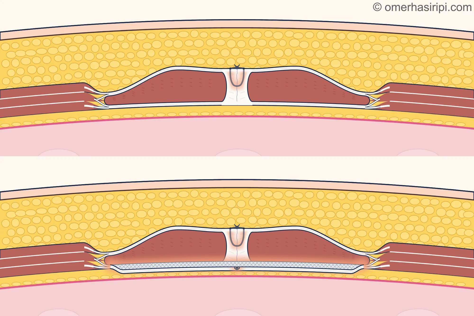
Enlarge
Swipe the drawing right or left to see the details. You can use the arrow keys on your keyboard.
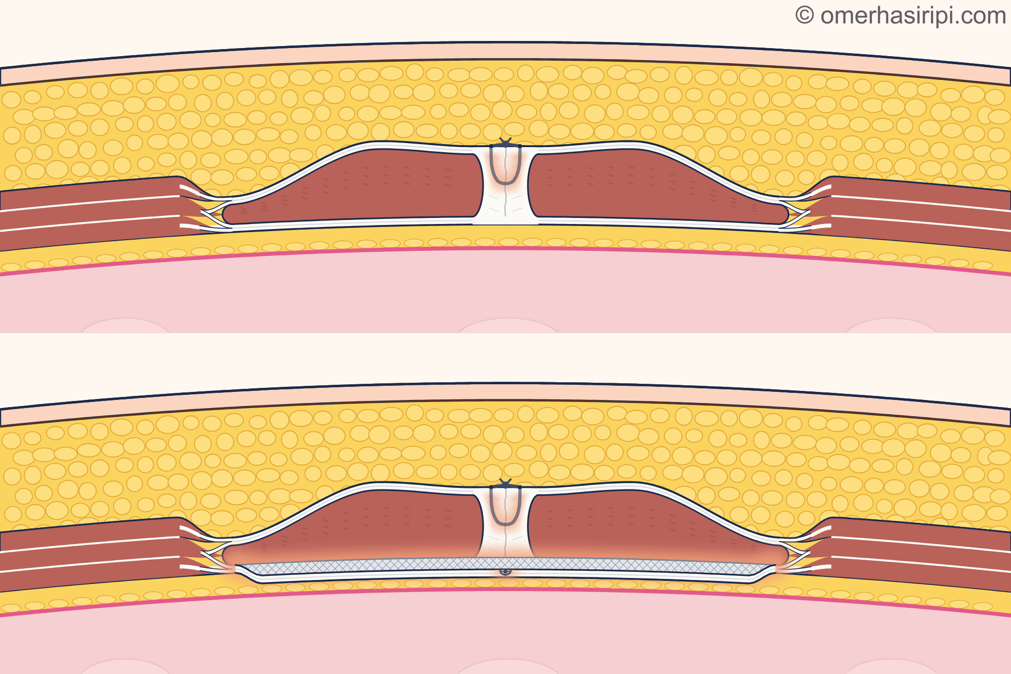
- 1 Repair with stitches
- 2 Repair with mesh
- 3 Stitch
- 4 Flat patch (mesh)
- 5 Long muscle of the front abdominal wall (rectus)
- 6 Firm layer where the muscle sheaths meet in the middle
- 7 Abdominal lining (peritoneum)
- 8 Fat tissue beneath the skin
Description of the drawing
Open repair of a midline incisional hernia, shown in cross-section through the front abdominal wall above the navel; in both drawings the front of the body is at the top and the inside of the abdomen is at the bottom. In the upper drawing, the opening is closed with stitches alone; in the lower drawing, a flat mesh is placed behind the long abdominal muscles in addition to the same stitches. The European Hernia Society recommends mesh repair for the midline incisional hernias covered by its guideline and placement of the mesh in the layer behind the muscles.[1, 9]
Results may vary from person to person with any surgical or interventional procedure. You are advised to discuss the procedure in detail with your doctor beforehand.
Illustrative drawing, not to scale. Prepared for this site.
What is recovery like after surgery and which symptoms should you look out for?
Pain and limits on movement vary from person to person; increase your activity as much as feels comfortable[1, 9]. The EHS gives avoiding heavy lifting and exercises that involve straining for four weeks after repair of the midline incisional hernias covered by its guidelines as expert opinion; this period may be insufficient for complex repairs[1]. The American College of Surgeons recommends not lifting anything heavier than about 4.5 kg and avoiding strenuous activity for at least six weeks[9]. These periods may vary depending on your operation; follow the surgical instructions you are given[1, 9]. Your return to work depends on the work you do and the extent of the operation; discuss a suitable timeframe with your surgeon[5, 9]. Wash your hands before and after touching the area around the wound, and follow your surgeon’s instructions for dressings and bathing[1, 9]. A hernia may recur even years later[9]. If abdominal pain starts suddenly or is severe after surgery, if you cannot pass either wind or stool, or if you cannot breathe, call 112 or go to the nearest emergency department[12]. If you have any of the symptoms in the “When is it an emergency?” section, follow the instructions there. If you have no emergency symptoms but your pain is increasing or does not go away, you have abdominal cramps, your temperature rises above 38 °C, you feel cold and shiver along with a high temperature, you vomit repeatedly, your abdomen swells, there is swelling, redness, bleeding or a bad smell at the wound, or discharge from the wound increases, contact your surgeon[9]. Also contact your surgeon if you have not passed stool within three days after surgery[9].
What should you do if you develop a hernia during pregnancy or around a stoma?
According to the EHS patient leaflet, planned repair of an umbilical or epigastric hernia during pregnancy can often be postponed until after childbirth. However, if the hernia suddenly becomes painful, does not go back in, the skin over it becomes red and hurts when touched, or you develop nausea or vomiting, seek surgical help immediately; if the blood supply to the tissue in the hernia is impaired, emergency surgery is needed[5]. For hernias around a stoma, the EHS 2023 rapid guideline addresses hernia prevention only in patients who are to have a permanent end colostomy (bringing the end of the large bowel out through the abdominal wall); it makes no recommendations for treating a hernia that has already developed[13].
Which department treats abdominal wall hernias?
For assessment and surgery of incisional and epigastric hernias, patients can attend the general surgery department[1, 5]. If you have noticed a new swelling, you can also see your family doctor for an initial examination[10].
Sources
16
- Sanders DL, Pawlak MM, Simons MP, Aufenacker T, Balla A, Berger C, et al. Midline incisional hernia guidelines: the European Hernia Society. Br J Surg. 2023;110:1732–1768. doi: 10.1093/bjs/znad284. PubMed: 37727928.
- Deerenberg EB, Henriksen NA, Antoniou GA, Antoniou SA, Bramer WM, Fischer JP, et al. Updated guideline for closure of abdominal wall incisions from the European and American Hernia Societies. Br J Surg. 2022. doi: 10.1093/bjs/znac302. PubMed: 36026550.
- Henriksen NA, Nazari T, Simons MP, Hope W, Montgomery A. Guidelines for Treatment of Umbilical and Epigastric Hernias From the European and Americas Hernia Societies–A Web-Based Survey on Surgeons’ Opinion. J Abdom Wall Surg. 2022;1:10260. Supplementary file: 2019 consensus voting text for the EHS/AHS umbilical and epigastric hernia recommendations; it is not the final guideline text. doi: 10.3389/jaws.2022.10260. PubMed: 38314164.
- European Hernia Society (EHS). Romanian translation and adaptation of the umbilical and epigastric hernia guideline. Translated by: Ioana Florea; based on the 2020 guideline. Text: europeanherniasociety.eu.
- European Hernia Society (EHS). Umbilical and epigastric hernia: information for patients. February 2020. Text: europeanherniasociety.eu.
- Hernández-Granados P, Henriksen NA, Berrevoet F, Cuccurullo D, López-Cano M, Nienhuijs S, et al. European Hernia Society guidelines on management of rectus diastasis. Br J Surg. 2021. doi: 10.1093/bjs/znab128. PubMed: 34595502.
- Hanzalova I, Schäfer M, Demartines N, Clerc D. Spigelian hernia: current approaches to surgical treatment, review. Hernia. 2022. doi: 10.1007/s10029-021-02511-8. PubMed: 34665343.
- van Steensel S, Bloemen A, van den Hil LCL, van den Bos J, Kleinrensink GJ, Bouvy ND. Pitfalls and clinical recommendations for the primary lumbar hernia based on a systematic review of the literature. Hernia. 2019. doi: 10.1007/s10029-018-1834-9. PubMed: 30315438.
- American College of Surgeons (ACS). Ventral Hernia Repair: patient information page. Updated: 2024. Text: www.facs.org.
- NHS. Hernia: patient information page. Last reviewed: 19 May 2026. Text: www.nhs.uk.
- NHS. Umbilical hernia: patient information page. Last reviewed: 24 November 2025. Text: www.nhs.uk.
- NHS. Stomach ache: patient information page. Last reviewed: 26 May 2023; the next review date (26 May 2026) has passed. Accessed: 22 October 2026. Text: www.nhs.uk.
- Stabilini C, Muysoms FE, Tzanis AA, Rossi L, Koutsiouroumpa O, Mavridis D, et al. EHS rapid guideline on parastomal hernia prevention. J Abdom Wall Surg. 2023. doi: 10.3389/jaws.2023.11549. PubMed: 38312414.
- NHS. Sudden confusion (delirium): patient information page. Last reviewed: 28 May 2024. Text: www.nhs.uk.
- OpenStax. Anatomy and Physiology 2e. 2022. Section 23.1: Overview of the Digestive System; full text accessed 28 October 2026. Text: openstax.org.
- Birindelli A, Sartelli M, Di Saverio S, Coccolini F, Ansaloni L, van Ramshorst GH, et al. 2017 update of the WSES guidelines for emergency repair of complicated abdominal wall hernias. World J Emerg Surg. 2017;12:37. doi: 10.1186/s13017-017-0149-y. PubMed: 28804507.
Medical review of the Turkish original: Op. Dr. Ömer Seyfullah Hasıripi, . This page is a translation checked by AI reviewers; the Turkish version prevails.
This page is for general information; it does not replace an examination. See a doctor about your symptoms.