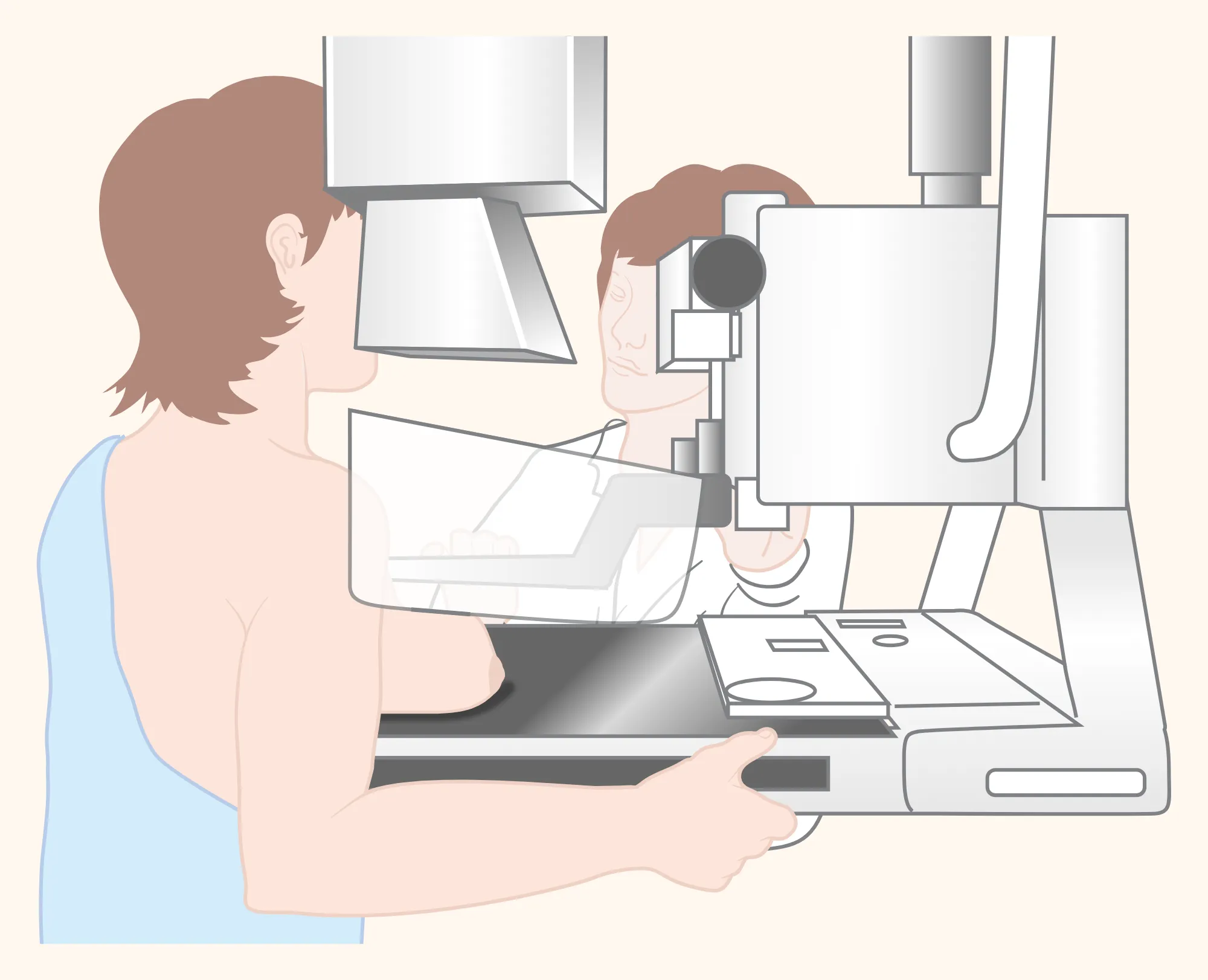
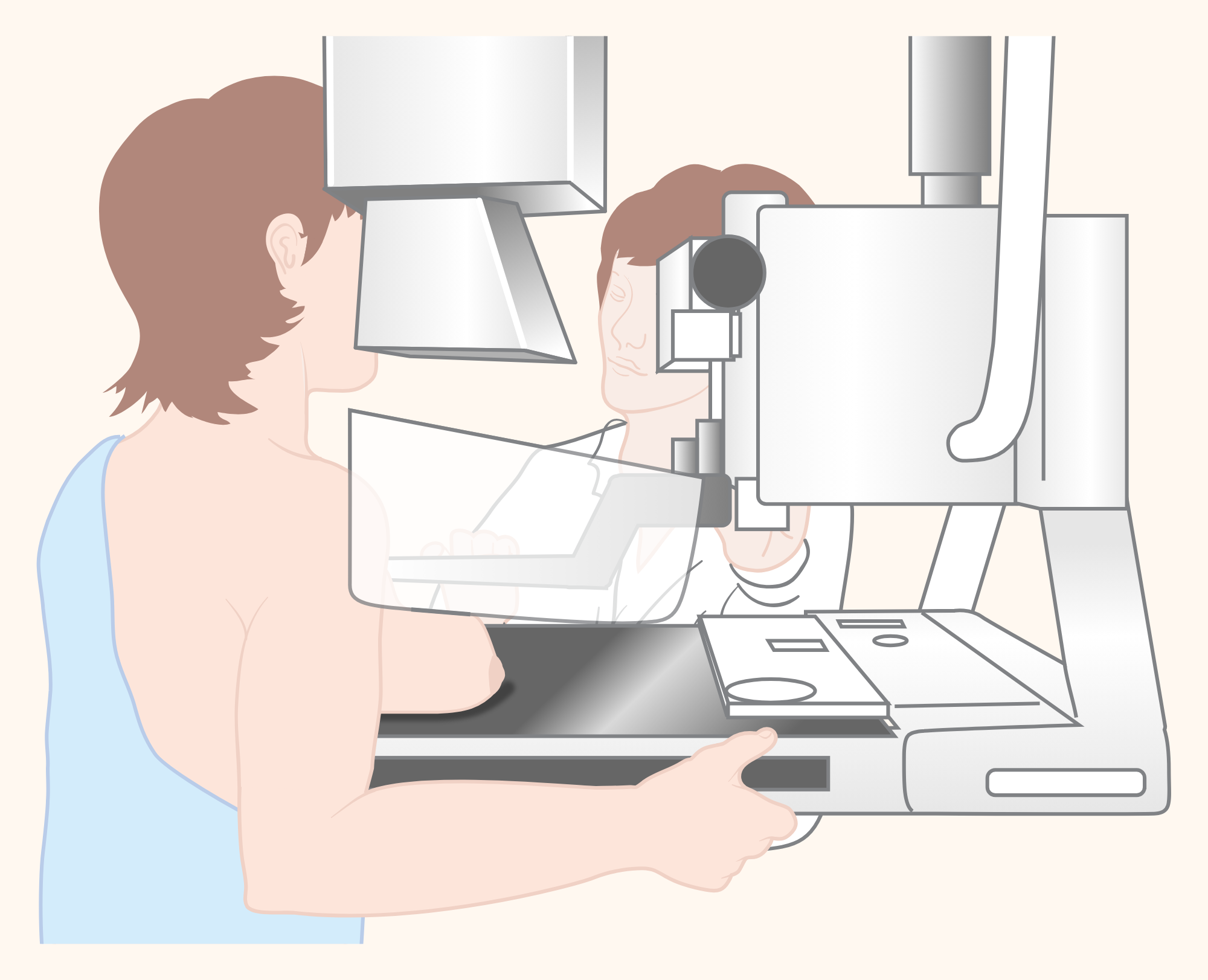

How is a mammogram done, and how many minutes does it take?
A mammogram is an image of the breast taken using low-dose X-rays[1]. During the examination, the breast is placed on a flat surface and gently compressed with a transparent plate[1]. The whole appointment takes about 30 minutes; the imaging takes a few minutes[1, 3].
A mammogram is often uncomfortable and may be painful for some people; the discomfort should not last long[2, 3]. Tell the technician if you have pain; if the discomfort is significant, less compression is used[1].
How do you prepare for a mammogram?
On the day of the examination, do not apply deodorant, powder or lotion to your armpits or breasts; they may look like calcium spots on the image[1]. Ask when your result will be ready; do not assume it is normal because you have not heard anything[1].
Is a mammogram harmful?
The radiation dose from a two-dimensional digital screening mammogram is about 0.28 millisieverts (mSv), equivalent to the average natural background radiation received in the US over about 34 days; this value can vary greatly depending on body size and the machine[4]. In a diagnostic mammogram taken because of a change, the dose is higher because images are taken from more angles[2]. Additional radiation always carries a small risk of cancer, but because the radiation used in imaging is low, the benefit of an accurate diagnosis is much greater[1].
How is a breast ultrasound performed, and why is it requested?
A breast ultrasound is performed by applying gel to the skin and sending sound waves through a small probe; no radiation is used and it should not be painful[5].
An ultrasound is requested to find out what a lump that can be felt, bloody or clear nipple discharge that occurs on its own, or a finding on a mammogram or MRI is[5]. However, many cancers do not show up on ultrasound; for example, DCIS (ductal carcinoma in situ), an early type of cancer, is usually not visible[5].
What is a breast MRI, and why is it requested?
A breast MRI creates images using a powerful magnet and radio waves; it does not use X-rays[6]. In most breast MRI scans, a medicine (contrast agent) is given through a vein during the scan[6]. If you have a device or metal in your body, tell the technician; if your kidneys are not working well or you are on dialysis, tell your doctor and the centre performing the scan[6]. MRI does not replace a mammogram or ultrasound; it may be used additionally to screen women at high risk of breast cancer, assess the extent of a cancer that has been diagnosed or investigate a finding on a mammogram[6]. On MRI, harmless tissue may also take up the medicine and appear bright, and may require an additional ultrasound or biopsy[6].
Ultrasound or mammogram: which is done first?
For a lump that can be felt, the first test is ultrasound in women under 30 and a diagnostic mammogram in women aged 40 and over; ultrasound is also performed for the lump[7, 8]. At ages 30–39, the British guidance starts with ultrasound, while the American guideline says that either ultrasound or mammography may be used first[7, 8]. According to the British guidance, ultrasound is also the first test during pregnancy and breastfeeding[8]; according to the American College of Radiology, during pregnancy, breast ultrasound is usually appropriate at any age for pain in one area of the breast or a lump that can be felt, with a diagnostic mammogram also usually appropriate at age 30 and over[23]; during breastfeeding, the American guideline recommends ultrasound as the first test for those under 30[24]. The approach to a lump that can be felt is described on the Breast lump page.
What does BI-RADS mean?
BI-RADS is a standard classification used by radiologists in mammogram, ultrasound and MRI reports[9].
- BI-RADS 0: The assessment is incomplete; additional imaging or comparison with older images is needed[9, 11].
- BI-RADS 1: The result is negative; there is no sign of cancer[9, 10].
- BI-RADS 2: There is a benign finding, such as a cyst[9, 10].
- BI-RADS 3: The finding is probably benign; the chance of cancer is at most 2%, and a follow-up check after 6 months is recommended[9, 10].
- BI-RADS 4: There is a suspicious finding; a biopsy is recommended (according to the US National Cancer Institute, “a biopsy may be needed”)[2, 9].
- BI-RADS 5: The chance of cancer is high (at least 95%); a biopsy is needed[2, 9, 10].
- BI-RADS 6: Cancer has already been diagnosed by biopsy[9, 11].
Routine screening is recommended for BI-RADS 1 and 2[10]. If there is a clearly suspicious finding on examination, normal imaging should not rule out a biopsy; surgical assessment or biopsy may be recommended even if the report is BI-RADS 1 or 2[7, 12].
What does BI-RADS 3 mean, and what happens next?
If a BI-RADS 3 finding does not change, it is usually monitored for 2 years; if it shrinks or disappears, it is assessed as BI-RADS 2 or 1 and follow-up may end sooner[12, 13]. According to the American College of Radiology’s 2022 guideline, imaging follow-up may be appropriate for a lump that can be felt in someone aged 40 or over and appears probably benign on a mammogram or ultrasound; but if the lump is new on imaging or its volume or every diameter has increased by more than 20% in 6 months, it is considered suspicious and biopsy is recommended[7]. The Royal College of Radiologists’ 2025 guidance states something different: patients with a lump that can be felt and an indeterminate or probably benign finding on ultrasound (U3) should also have a biopsy[8].
What do BI-RADS 4A, 4B and 4C mean?
In the BI-RADS 4 subgroups, the chance of cancer is more than 2% and at most 10% for 4A, more than 10% and at most 50% for 4B, and more than 50% and less than 95% for 4C[9, 14].
What does “dense breast” mean in the report?
A mammogram report also records breast tissue density as one of four grades[9]. Dense breasts are not a disease; about half of women over 40 have dense breasts[15]. Dense breast tissue may also increase the risk of breast cancer somewhat[15]; the Turkish Ministry of Health lists it among the risk factors for breast cancer[27]. In addition, a mammogram may miss cancer more easily in dense breasts; discuss what this means for you with your doctor[2, 15].
Why is a breast biopsy requested, and how is it performed?
Examination and imaging cannot always distinguish whether a finding is benign or cancer[16]. The aim of a needle biopsy is to take a small sample of the finding, not the whole finding[16]. A radiologist most often performs the biopsy with a needle under imaging guidance[16]. The needle is guided by ultrasound, mammography or MRI; a biopsy performed using mammography may be called a stereotactic biopsy[17].
- Fine needle aspiration: fluid or cells are taken with a very fine needle[16, 17].
- Core needle biopsy: a piece of tissue is taken with each insertion using a thicker, hollow needle[16, 17].
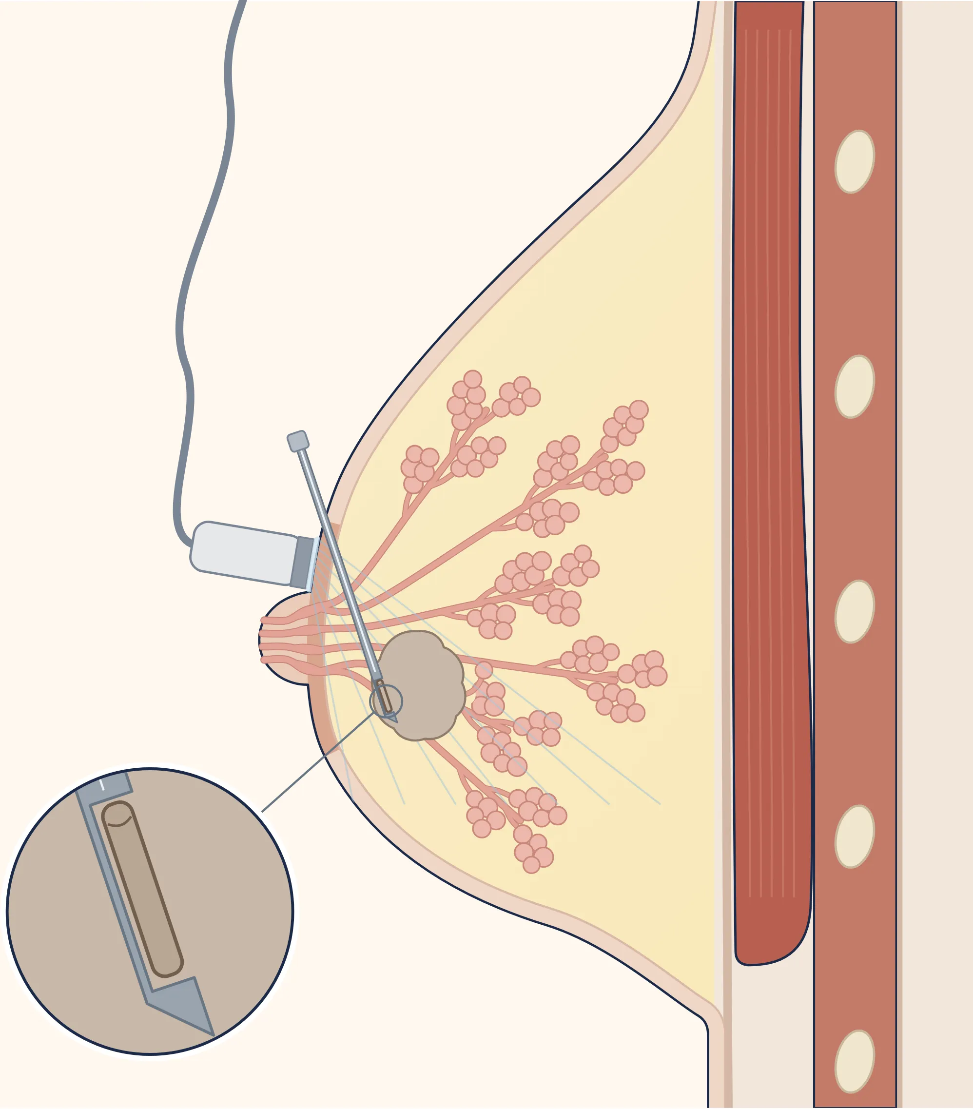
Enlarge
Swipe the drawing right or left to see the details. You can use the arrow keys on your keyboard.
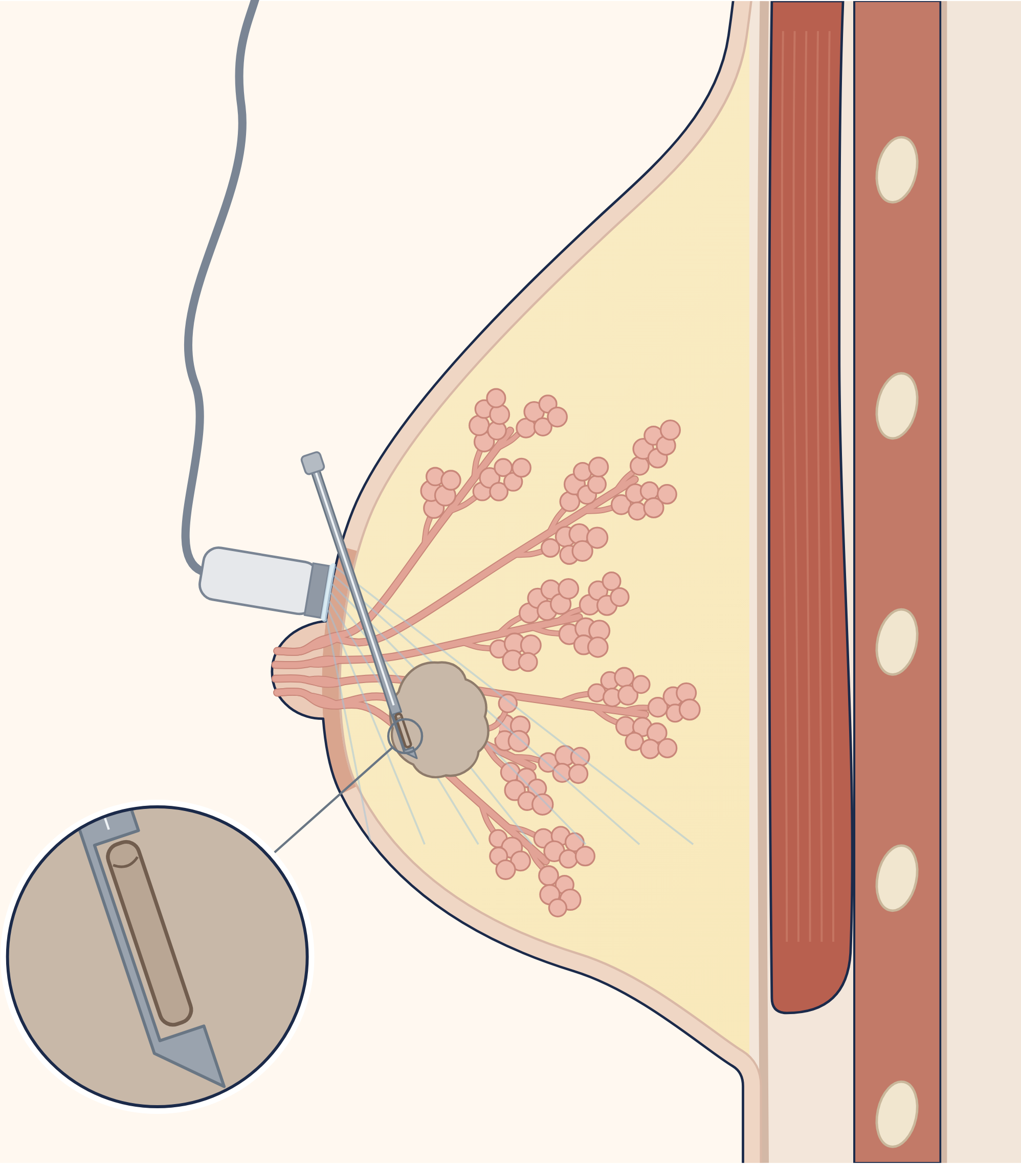
- 1 Ultrasound head (probe)
- 2 Gel applied to the skin
- 3 Thick, hollow biopsy needle
- 4 Finding being sampled
- 5 Small piece of tissue taken inside the needle
- 6 Chest muscle
Description of the drawing
Ultrasound-guided core needle biopsy: the radiologist sees the finding with the ultrasound probe and advances the thick, hollow needle through the skin to the finding. With each insertion, a small piece is taken, not the whole finding.[16, 17]
Results may vary from person to person with any surgical or interventional procedure. You are advised to discuss the procedure in detail with your doctor beforehand.
Illustrative drawing, not to scale. Source: Cancer Research UK / Wikimedia Commons, CC BY-SA 4.0. The English text and pointer lines were removed; the breast outline and position of the nipple in the source were retained, with only the lower end redrawn to complete the fold beneath the breast; the skin, nipple and surrounding ring, milk glands, ducts, chest muscle and ribs were redrawn, and the fatty tissue was coloured; the finding, ultrasound probe, gel, sound lines, biopsy needle, tissue sample inside the needle and enlarged view of the needle tip were added; a small sac beside the finding was moved slightly; numbered labels were added. This adaptation is also licensed under CC BY-SA 4.0.

Does a breast biopsy hurt, and how long does it take?
Before an ultrasound-guided core needle biopsy, the skin and breast tissue are usually numbed with a local anaesthetic[16, 28]. For fine needle aspiration, a local anaesthetic is most often not needed[19]. If you are allergic to a local anaesthetic medicine, contrast agent or another medicine, tell your doctor and inform the team performing the procedure as soon as possible when your appointment is arranged[6, 16, 28]. You can ask the imaging team performing the procedure how your biopsy will be done. You are awake during the procedure; many women say they feel little pain[16]. An ultrasound-guided biopsy is usually completed within an hour[16]. A small metal marker (clip) may be left at the biopsy site so it can be found later[16, 17]. The clip can remain in the breast and does not usually cause pain[16]. Clips may contain nickel; if you are allergic or sensitive to nickel, tell the team performing the procedure before the clip is placed[30].
Is there pain after a breast biopsy, and what are the risks?
There may be swelling and bruising after a biopsy; temporary bruising is normal and your doctor may recommend applying something cold[16]. If you have pain, you can take a mild painkiller such as paracetamol; it is best to avoid ibuprofen or painkillers containing aspirin because they can cause bruising in the breast[28]. Your doctor will give you detailed care instructions; how long to avoid strenuous activities varies by source: at least 24 hours[16] or the rest of the day of the procedure[28].
Serious problems requiring intervention are rare[19]. Bleeding and a collection of blood (haematoma) can occur at the biopsy site[16]. In a biopsy of tissue deep in the breast, it is very rare for the needle to pass through the chest wall and collapse the lung[16]. According to RadiologyInfo, the chance of an infection requiring antibiotics after an ultrasound-guided breast biopsy appears to be less than 1 in 1,000[16]. A biopsy can sometimes miss the finding or may not provide a definite answer; if the diagnosis remains uncertain, a surgical biopsy is usually needed[16]. The next step after biopsy is determined by assessing the imaging and pathology findings together[16, 21].
Details of emergency symptoms
Severe difficulty breathing means gasping, feeling as though you are choking or being unable to speak[29]. Blue, grey, pale or mottled colouring may be easier to see on the palms or soles of people with darker skin[29, 33]. Sepsis is a severe response to an infection that can be life-threatening; its other symptoms are described on the mastitis page[33].
Bleeding requiring emergency care is bleeding at the biopsy site that you cannot stop, or bright red, spurting bleeding that is difficult to stop[31]. Redness at the biopsy site may be harder to see on darker skin[32].
What should you do if the biopsy site bleeds?
If the centre that performed the procedure gave you written care instructions, follow them for daily care[16, 28]; but do not delay getting help for situations in the “When is it an emergency?” box that require 112 or an emergency department. Bruising at the biopsy site is expected and should decrease within 7–10 days; bleeding or swelling is rare: if it occurs, remove the dressing and press firmly on the biopsy area with the palm of your hand using a paper tissue or paper towel for 10 minutes; if bleeding or swelling continues after 15 minutes, go to the nearest emergency department[28]. If blood is spurting, do not wait for these periods: call 112 or go to the nearest emergency department[31].
What should you do if your mammogram is normal or if you develop a new symptom during pregnancy or breastfeeding?
Even if your mammogram is normal, see a specialist doctor if you have a suspicious symptom; a normal result does not guarantee that you do not have cancer or will not develop it in the future[22]. The “When is it an emergency?” box on the Breast lump page lists the symptoms for which you should see a doctor the same day.
If you notice a new lump in your breast, show it to a doctor without waiting for it to go away[20]; natural breast changes during pregnancy can make findings that can be felt harder to notice and may play a role in delayed diagnosis[23]. According to the American guideline, a noticeable lump that persists for two weeks or longer during pregnancy should be assessed[23]; this does not mean you should wait two weeks.
If you notice a new change in your breast that you can feel, signs of inflammation that do not go away or suspicious nipple discharge while breastfeeding, see a doctor; these should be investigated with imaging and, if necessary, a biopsy should be performed[24]. The Mastitis and breast abscess page explains when to see a doctor for breast pain, redness, warmth or swelling while breastfeeding.
What if I take blood thinners?
If you take aspirin, blood thinners or herbal supplements, tell your doctor and the team performing the procedure before the biopsy; your doctor may recommend stopping some of them to reduce the risk of bleeding[16, 28]. When your appointment is arranged, ask the team performing the procedure whether you should stop your medicine[28]. You may also need to stop aspirin or blood thinners for a short time after the procedure; ask the team that performed it about this before going home, and if you have a question about your medicines afterwards, call the centre where the procedure was performed[28].
How many days does it take to get a breast biopsy result?
A pathologist examines the sample taken and makes the definitive diagnosis[16]. You may need to wait about a week for the biopsy result[20]. Ask your doctor when the result will be ready and how you will be told, and make sure your results appointment has been arranged; if you have not heard within the expected time, it is fine to ask again[17, 28].
What does “B3” mean in a biopsy result?
The result may also be “B3” (a lesion of uncertain malignant potential or a high-risk lesion)[19]. B3 is a group of different findings for which the chance of cancer being present alongside them varies according to the type of finding[19]. B3 is a biopsy result; it is not the same as BI-RADS 3 in an imaging report[9, 19]. BI-RADS 3 is not assigned directly from a screening mammogram; it is assigned only after a diagnostic examination[10, 12].
Which department should you visit after a mammogram result?
The radiologist sends the report to the doctor who requested the test; that doctor most often explains the result, although the radiologist may sometimes explain it too[1, 5]. Ask the imaging centre whether you can access your report online[9]. According to the Turkish Ministry of Health’s cancer screening guideline, a person whose mammogram report shows a problem is referred to a general surgeon for a more detailed examination to reach a definitive diagnosis[22].
Can you have a mammogram during pregnancy and breastfeeding?
Mammography is considered safe during pregnancy; the scattered radiation reaching the baby is negligible[23]. In an ultrasound-guided biopsy, no radiation is used for guidance; if a marker (clip) is left at the biopsy site, a mammogram may be taken to confirm it is in the right place[16]. According to the American College of Radiology’s 2025 guideline, ultrasound-guided breast procedures and, if necessary, core needle biopsy can be performed safely during pregnancy[23]. If there is a possibility that you are pregnant, always tell your doctor and the team performing the procedure[18]. MRI with medicine (contrast) is not recommended during pregnancy because the medicine crosses the placenta[23]. If a biopsy is needed during pregnancy, milk fistula (milk leaking through the biopsy tract) and the increased risk of bleeding should be discussed beforehand[23, 25].
You do not need to pause breastfeeding or discard expressed milk after a mammogram[24]. After MRI medicine, manufacturers recommend not breastfeeding for 24–48 hours; the American College of Radiology, however, does not consider a pause necessary[6, 24]. A core needle biopsy can also be performed safely during breastfeeding when needed; rare risks such as milk fistula and bleeding should be discussed beforehand, and stopping breastfeeding before the biopsy is not recommended[24, 25].
I have a breast implant (prosthesis). Can I have a mammogram?
According to the US National Cancer Institute, yes: women with breast implants should continue to have mammograms; however, for most women whose breast has been removed (mastectomy) and reconstructed with an implant, a mammogram of the reconstructed breast is usually not needed[2]. A breast implant (prosthesis) can hide some breast tissue; tell the centre, as special images can be taken with the implant pushed aside[2]. The risk of compression and needle biopsy damaging the implant is small[8].
How is breast imaging performed in men?
Most breast problems in men are benign; the most common cause is gynaecomastia (enlargement of breast tissue). If typical gynaecomastia is seen on examination, imaging is not recommended[26]. For an uncertain lump in men, the American guideline (2018) starts with ultrasound in those younger than 25 and mammography in those aged 25 and over; if cancer is suspected on examination, mammography or three-dimensional mammography, with ultrasound as a complement, is usually appropriate at any age[26]. The British guidance (2025) recommends ultrasound for those younger than 40; ultrasound or mammography may be used at age 40 and over[8].
Sources
33
- RadiologyInfo.org (patient website of the American College of Radiology and the Radiological Society of North America). Mammography (Mammogram) (mammography): patient information page. Last reviewed: 4 February 2025. Accessed: 5 October 2026. Text: www.radiologyinfo.org.
- National Cancer Institute (NCI, US National Cancer Institute). Mammograms (mammography): patient information page. Last updated: 2 December 2025. Accessed: 5 October 2026. Text: www.cancer.gov.
- NHS (National Health Service in England). What happens at your breast screening appointment (what happens at a breast screening appointment): patient information page. Last reviewed: 9 October 2025. Accessed: 5 October 2026. Text: www.nhs.uk.
- RadiologyInfo.org. Radiation Dose (radiation dose in X-ray and CT examinations): patient information page. Last reviewed: 15 April 2025. Accessed: 5 October 2026. Text: www.radiologyinfo.org.
- RadiologyInfo.org. Breast Ultrasound (breast ultrasound): patient information page. Last reviewed: 15 June 2026. Accessed: 5 October 2026. Text: www.radiologyinfo.org.
- RadiologyInfo.org. Breast MRI (breast MRI): patient information page. Last reviewed: 15 June 2026. Accessed: 5 October 2026. Text: www.radiologyinfo.org.
- Klein KA, Kocher M, Lourenco AP, Niell BL, Bennett DL, Chetlen A, et al. ACR Appropriateness Criteria® Palpable Breast Masses: 2022 Update (2022 update, published in 2023). J Am Coll Radiol. 2023;20(5S):S146–S163. doi: 10.1016/j.jacr.2023.02.013. PubMed: 37236740.
- The Royal College of Radiologists. Guidance on screening and symptomatic breast imaging. 5th edition, October 2025. (Endorsed by the Association of Breast Surgery.) Text: associationofbreastsurgery.org.uk.
- RadiologyInfo.org. How to Read Your Breast Imaging Report using BI-RADS (breast imaging report and BI-RADS): patient information page. Last reviewed: 9 September 2024. Accessed: 5 October 2026. (The BI-RADS book itself could not be read because it is paid; the category definitions and rates were taken from this page and sources 10–14.) Text: www.radiologyinfo.org.
- Kaplan E, Aydemir AB. How Does ACR BI-RADS® v2025 Change the Radiologist's Approach? A Practical Guide Across Mammography, Ultrasound, and MRI: A Narrative Review. Diagnostics (Basel). 2026;16(13):2135. (A review describing the BI-RADS 2025 edition; not the edition itself.) doi: 10.3390/diagnostics16132135. PubMed: 42449916.
- American College of Radiology (American College of Radiology). ACR BI-RADS® v2025 Manual – What's New? November 2025. (A free document summarising the changes in the new edition; not the edition itself.) Text: edge.sitecorecloud.io.
- Kang BJ. Breast Imaging Reporting and Data System v2025: Key Updates in Mammography. J Korean Soc Radiol. 2026;87(3):437–459. (A review describing the BI-RADS 2025 edition; not the edition itself.) doi: 10.3348/jksr.2026.0028. PubMed: 42306164.
- Kim JY. Breast Imaging Reporting and Data System v2025: Key Updates in MRI. J Korean Soc Radiol. 2026;87(3):473–484. (A review describing the BI-RADS 2025 edition; not the edition itself.) doi: 10.3348/jksr.2026.0042. PubMed: 42306163.
- Elezaby M, Li G, Bhargavan-Chatfield M, Burnside ES, DeMartini WB. ACR BI-RADS Assessment Category 4 Subdivisions in Diagnostic Mammography: Utilization and Outcomes in the National Mammography Database. Radiology. 2018;287(2):416–422. (Reports the 4A–4C definitions from the 5th edition of BI-RADS.) doi: 10.1148/radiol.2017170770. PubMed: 29315061.
- RadiologyInfo.org. Dense Breasts (dense breast tissue): patient information page. Last reviewed: 11 March 2024. Accessed: 5 October 2026. Text: www.radiologyinfo.org.
- RadiologyInfo.org. Ultrasound-Guided Breast Biopsy (ultrasound-guided breast biopsy): patient information page. Last reviewed: 1 June 2026. Accessed: 5 October 2026. Text: www.radiologyinfo.org.
- National Cancer Institute (NCI, US National Cancer Institute). How Breast Cancer Is Diagnosed (how breast cancer is diagnosed): patient information page. Published: 2 December 2025. Accessed: 5 October 2026. Text: www.cancer.gov.
- RadiologyInfo.org. Stereotactic Breast Biopsy (mammography-guided breast biopsy): patient information page. Last reviewed: 11 March 2024. Accessed: 5 October 2026. Text: www.radiologyinfo.org.
- Sanderink WBG, Camps-Herrero J, Athanasiou A, Couto HL, Mehta K, Palak Bhavesh Thakkar P, et al. Image-guided biopsy of breast lesions — when to use what biopsy technique. Insights Imaging. 2025;16(1):208. (Consensus report by eight experts. Participants were selected after a meeting between the study organisers and the sponsor; a medical device company (BD) supported the meeting and, according to the article, did not participate in the discussion.) doi: 10.1186/s13244-025-02084-5. PubMed: 40996584.
- NHS (National Health Service in England). Breast lumps (breast lump): patient information page. Last reviewed: 28 June 2023 (the page's stated review date of 28 June 2026 has passed). Accessed: 5 October 2026. Text: www.nhs.uk.
- Yincharoen P, Sharma CP, Tawanwongsri W. Radiologic–Pathologic Discordance After Image-Guided Breast Biopsy: A Systematic Review of Prevalence and Outcomes. Med Sci (Basel). 2026;14(2):229. doi: 10.3390/medsci14020229. PubMed: 42201021.
- Turkish Ministry of Health. Aile Sağlığı Merkezlerinde Kanser Tarama Kılavuzu. Electronically signed PDF on the Ministry’s dosyaism.saglik.gov.tr server (no date printed in the document; PDF file created on 30 June 2021).
- Salkowski LR, Lewin AA, Weinstein SP, Bartell S, Birsner ML, Borges VF, et al. ACR Appropriateness Criteria® Breast Imaging During Pregnancy. J Am Coll Radiol. 2025;22(11S):S492–S507. doi: 10.1016/j.jacr.2025.08.045. PubMed: 41193040.
- Dogan BE, Salkowski LR, Weinstein SP, Bartell S, Chikarmane S, Dibble EH, et al. ACR Appropriateness Criteria® Breast Imaging During Lactation. J Am Coll Radiol. 2026;23(8):1768–1785. doi: 10.1016/j.jacr.2026.03.002. PubMed: 41885663.
- Mitchell KB, Johnson HM, Eglash A; Academy of Breastfeeding Medicine. ABM Clinical Protocol #30: Breast Masses, Breast Complaints, and Diagnostic Breast Imaging in the Lactating Woman. Breastfeed Med. 2019;14(4):208–214. doi: 10.1089/bfm.2019.29124.kjm. PubMed: 30892931.
- Niell BL, Lourenco AP, Moy L, Baron P, Didwania AD, diFlorio-Alexander RM, et al. ACR Appropriateness Criteria® Evaluation of the Symptomatic Male Breast. J Am Coll Radiol. 2018;15(11S):S313–S320. doi: 10.1016/j.jacr.2018.09.017. PubMed: 30392600.
- Turkish Ministry of Health, General Directorate of Public Health, Department of Cancer. 1-31 Ekim Meme Kanseri Farkındalık Ayı. Page footer: Copyright © 2025. Accessed: 5 October 2026. Text: hsgm.saglik.gov.tr.
- Guy's and St Thomas' NHS Foundation Trust (London). Ultrasound-guided core biopsy of the breast (ultrasound-guided breast core needle biopsy): patient information page, 3322/VER7. Last reviewed: October 2026. Accessed: 5 October 2026. (Patient page from a single NHS hospital.) Text: www.guysandstthomas.nhs.uk.
- NHS (National Health Service in England). Shortness of breath (shortness of breath): patient information page. Last reviewed: 30 January 2024. Accessed: 5 October 2026. (General page on shortness of breath; not specific to biopsy.) For England only: the directions on the page (999, A&E, NHS 111) belong to the healthcare system in England. Text: www.nhs.uk.
- Belfast Health and Social Care Trust (Northern Ireland). Patient Information Leaflet for having a Breast Marker Clip Inserted (insertion of a breast marker clip): patient information leaflet, version 4.0. Published: 23 January 2025; planned review: January 2027. Accessed: 6 October 2026. (Leaflet from a single hospital group.) (Written for a separate clip insertion procedure; used only for information about the clip material and nickel.) Text: belfasttrust.hscni.net.
- NHS (National Health Service in England). Cuts and grazes (cuts and grazes): patient information page. Last reviewed: 2 April 2026. Accessed: 6 October 2026. (General first aid advice for cuts; not specific to biopsy.) For England only: the directions on the page (999, A&E, NHS 111) belong to the healthcare system in England. Text: www.nhs.uk.
- NHS (National Health Service in England). Biopsy (biopsy): patient information page. Last reviewed: 11 June 2025 (next review: 11 June 2028). Accessed: 6 October 2026. (General biopsy page; not specific to breast biopsy.) For England only: the direction on the page (NHS 111) belongs to the healthcare system in England. Text: www.nhs.uk.
- NHS (National Health Service in England). Sepsis: patient information page. Last reviewed: 14 May 2026 (next review: 14 May 2029). Accessed: 6 October 2026. (General sepsis page; not specific to biopsy.) The NHS sepsis page also lists a very high or very low body temperature, feeling hot or cold to the touch, and shivering at the 999 level. By the doctor's decision, the box uses the same-day level for a high temperature after biopsy (including feeling hot to the touch) and feeling cold and shivering with or without a fever, as does the site's Mastitis page (tell the team that performed the procedure immediately or see a doctor the same day; for a high temperature, see the NHS biopsy page, source 32); a very low body temperature, feeling cold to the touch and suspected sepsis are at the 112 level. For England only: the directions on the page (999, A&E, NHS 111) belong to the healthcare system in England. Text: www.nhs.uk.
Medical review of the Turkish original: Op. Dr. Ömer Seyfullah Hasıripi, . This page is a translation checked by AI reviewers; the Turkish version prevails.
This page is for general information; it does not replace an examination. See a doctor about your symptoms.